. 2021 Jul 23;13(15):2420.
doi: 10.3390/polym13152420.

Counter-Acting Candida albicans- Staphylococcus aureus Mixed Biofilm on Titanium Implants Using Microbial Biosurfactants

Affiliations

Counter-Acting Candida albicans- Staphylococcus aureus Mixed Biofilm on Titanium Implants Using Microbial Biosurfactants

Erica Tambone et al. Polymers (Basel). .

Abstract

This study aimed to grow a fungal-bacterial mixed biofilm on medical-grade titanium and assess the ability of the biosurfactant R89 (R89BS) coating to inhibit biofilm formation. Coated titanium discs (TDs) were obtained by physical absorption of R89BS. Candida albicans-Staphylococcus aureus biofilm on TDs was grown in Yeast Nitrogen Base, supplemented with dextrose and fetal bovine serum, renewing growth medium every 24 h and incubating at 37 °C under agitation. The anti-biofilm activity was evaluated by quantifying total biomass, microbial metabolic activity and microbial viability at 24, 48, and 72 h on coated and uncoated TDs. Scanning electron microscopy was used to evaluate biofilm architecture. R89BS cytotoxicity on human primary osteoblasts was assayed on solutions at concentrations from 0 to 200 μg/mL and using eluates from coated TDs. Mixed biofilm was significantly inhibited by R89BS coating, with similar effects on biofilm biomass, cell metabolic activity and cell viability. A biofilm inhibition >90% was observed at 24 h. A lower but significant inhibition was still present at 48 h of incubation. Viability tests on primary osteoblasts showed no cytotoxicity of coated TDs. R89BS coating was effective in reducing C. albicans-S. aureus mixed biofilm on titanium surfaces and is a promising strategy to prevent dental implants microbial colonization.

Keywords: Candida albicans; Staphylococcus aureus; anti-biofilm coating; biosurfactants; cytotoxicity; dental implant; fungal-bacterial biofilm; mixed biofilm; peri-implantitis; scanning electron microscopy; titanium coating.


Conflict of interest statement

The authors declare no conflict of interest. The funders of the study and the companies that provided the titanium discs had no role in the design of the study; in the collection, analyses, or interpretation of data; in the writing of the manuscript, or in the decision to publish the results.

Figures

Figure 1
Figure 1
Biomass (a), metabolic activity (b) and cell viability (c) of C. albicans ATCC ®® 10231 and S. aureus ATCC ®® 6538 single- and multi-species biofilm formed on untreated titanium discs. Results were obtained by mean CV staining, MTT reduction assay and viable cell counting respectively. Single- and multi-species cultures were grown in YNBD + 10% FBS and were evaluated after 24, 48 and 72 h of incubation. The multi-species biofilm composition (d) was determined by viable cell counting on selective agar media (Mannitol Salt Agar, selective for Staphylococcus, and Sabouraud Chloramphenicol Agar, selective for fungal growth) at the same time-points (24 h: internal pie; 48 h: middle pie; 72 h: external pie). * p < 0.05.
Figure 2
Figure 2
Biomass (a), metabolic activity (b) and cell viability (c) of C. albicans-S. aureus multi-species biofilm formed on untreated control TDs (dark bars) and TDs coated with 4 mg/mL R89BS (light bars). Results were obtained by means of CV staining, MTT reduction assay and viable cell counting respectively, after 24, 48 and 72 h of incubation. The composition of the multi-species biofilm formed on R89BS-coated TDs (d) was determined by viable cell counting on selective agar media (Mannitol Salt Agar, selective for Staphylococcus, and Sabouraud Chloramphenicol Agar, selective for fungal growth) at the same time-points (24 h: internal pie; 48 h: middle pie; 72 h: external pie). Error bars represent standard deviation. * p < 0.05.
Figure 3
Figure 3
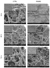
Architecture of C. albicans-S. aureus dual-species biofilm grown on uncoated control TDs (CTRL, panels (a,c,e)) and R89BS-coated TDs (R89BS, panels (b,d,f), after 24, 48 and 72 h of incubation. Representative images were obtained by scanning electron microscopy in high-vacuum mode. Areas free of adherent microorganisms in (b) are well representing the micro-morphology of both coated and uncoated titanium discs. Original magnification: 4000× (insets 500×).
Figure 4
Figure 4
Results of the eucaryotic cell viability MTT test: (a) hOBs viability data with R89BS added as aqueous solution at concentrations ranging from 200 to 0 µg/mL (Ctrl −); (b) Viability data obtained from hOBs cultured in the eluate from R89BS-coated discs (R89BS-TDs) and uncoated titanium discs (Ctrl −). Triton X-100 was used as positive control (Ctrl +). Error bars represent standard deviations. (* p < 0.05).

References

    1. Dewhirst F.E., Chen T., Izard J., Paster B.J., Tanner A.C.R., Yu W.-H., Lakshmanan A., Wade W.G. The Human Oral Microbiome. J. Bacteriol. 2010;192:5002–5017. doi: 10.1128/JB.00542-10. - DOI - PMC - PubMed
    1. Khurshid Z., Naseem M., Sheikh Z., Najeeb S., Shahab S., Zafar M.S. Oral antimicrobial peptides: Types and role in the oral cavity. Saudi Pharm. J. 2016;24:515–524. doi: 10.1016/j.jsps.2015.02.015. - DOI - PMC - PubMed
    1. Marsh P.D., Zaura E. Dental biofilm: Ecological interactions in health and disease. J. Clin. Periodontol. 2017;44:S12–S22. doi: 10.1111/jcpe.12679. - DOI - PubMed
    1. Montelongo-Jauregui D., Lopez-Ribot J.L. Candida Interactions with the Oral Bacterial Microbiota. J. Fungi. 2018;4:122. doi: 10.3390/jof4040122. - DOI - PMC - PubMed
    1. Negrini T.D.C., Koo H., Arthur R.A. Candida–Bacterial Biofilms and Host–Microbe Interactions in Oral Diseases. Adv. Exp. Med. Biol. 2019;1197:119–141. doi: 10.1007/978-3-030-28524-1_10. - DOI - PubMed

LinkOut - more resources