Review
. 2021 Jul 31;13(15):2540.
doi: 10.3390/polym13152540.

Nanomaterial-Based Dual-Emission Ratiometric Fluorescent Sensors for Biosensing and Cell Imaging

Affiliations
Review

Nanomaterial-Based Dual-Emission Ratiometric Fluorescent Sensors for Biosensing and Cell Imaging

Yanan Zhang et al. Polymers (Basel). .

Abstract

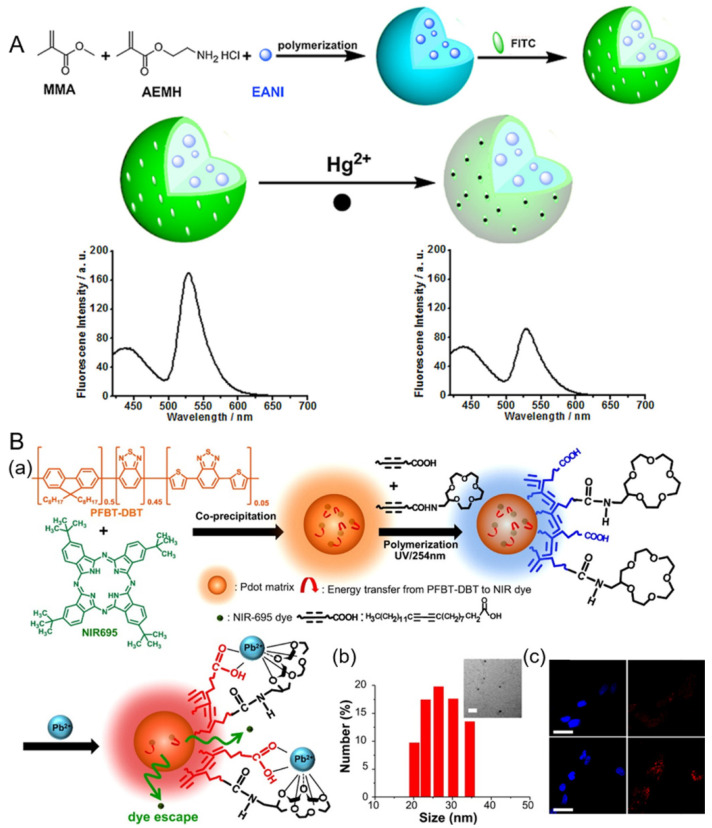
Owing to the unique optophysical properties of nanomaterials and their self-calibration characteristics, nanomaterial-based (e.g., polymer dots (Pdots) quantum dots (QDs), silicon nanorods (SiNRs), and gold nanoparticle (AuNPs), etc.) ratiometric fluorescent sensors play an essential role in numerous biosensing and cell imaging applications. The dual-emission ratiometric fluorescence technique has the function of effective internal referencing, thereby avoiding the influence of various analyte-independent confounding factors. The sensitivity and precision of the detection can therefore be greatly improved. In this review, the recent progress in nanomaterial-based dual-emission ratiometric fluorescent biosensors is systematically summarized. First, we introduce two general design approaches for dual-emission ratiometric fluorescent sensors, involving ratiometric fluorescence with changes of one response signal and two reversible signals. Then, some recent typical examples of nanomaterial-based dual-emission ratiometric fluorescent biosensors are illustrated in detail. Finally, probable challenges and future outlooks for dual-emission ratiometric fluorescent nanosensors for biosensing and cell imaging are rationally discussed.

Keywords: biosensing; cell imaging; nanomaterial; ratiometric fluorescent sensor.


Conflict of interest statement

The authors declare no conflict of interest.

Figures

Figure 1
Figure 1
The general categories for fabricating dual-emission ratiometric fluorescent sensors. Ratiometric fluorescence with one reference signal (A) or with two reversible signal changes (B). Reproduced from [1], with permission from Elsevier, 2019.
Figure 2
Figure 2
(A) Schematic description of (a) the structure of ps-po DNA (ps backbone: phosphorothioate linkage, which can bind to QDs due to their high affinity to cadmium; po backbone: phosphate linkage, which is the basic component of DNA.), (b) preparation of Rox-DNA functionalized CdZnTeS QDs, and (c) H2O2 detection based on Rox-DNA functionalized CdZnTeS QDs. Reproduced from [42], with permission from the American Chemical Society, 2017. (B) Representation of fluorescence detection of CHL through IFE of gold nanoparticles on RF-QDs. Reproduced from [36], with permission from the American Chemical Society, 2020. (C) Proposed mechanism of fluorescence sensing guanine by CDs-ZnCdTe QDs. Reproduced from [43], with permission from Elsevier, 2018.
Figure 3
Figure 3
(A) Schematic presentation of the preparation of SiND-DNA-Rox and its application for ratiometric detection of Hg2+. Reproduced from [23], with permission from the American Chemical Society, 2018. (B) (a) Schematic of Eu@SiNRs sensor, (b) TEM image of Eu@SiNRs, (c) fluorescence spectra of Eu@SiNRs in PBS buffers with different pH values under 405 nm excitation, (d) a Eu@SiNRs-based pH sensor for ratiometric measurements of cytoplasmic pH in live cells. Reproduced from [56], with permission from the American Chemical Society, 2017.
Figure 4
Figure 4
(A) Preparation of dual-labeled carbon nanodots (DLCDs). a: TTDDA, 220 °C, 3 h; b: FITC, RBITC, room temperature, overnight. Reproduced from [58], with permission from Wiley, 2012. (B) Dual-emission fluorescence sensing of Hg2+ based on a CNP–RhB nanohybrid system. Images of A549 cells after being incubated with 200 μL of CNP–RhB nanohybrid solution in the (ad) absence and (eh) presence of Hg2+; (a,e) bright-field images; (b,f) blue fluorescence field images; (c,g) red fluorescence field images; (d) the merged images (b,c); (h) the merged images (f,g). The scale bar is 20 μm. Reproduced from [59], with permission from the American Chemical Society, 2014. (C) AuNCs–CD sensor for ratiometric fluorescence imaging of intracellular Fe3+. Reproduced from [60], with permission from the American Chemical Society, 2016.
Figure 5
Figure 5
(A) Schematic of FRET probe for ratiometric imaging of intracellular telomerase. (B) Confocal images of HeLa, MCF-7, HepG2, and L-O2 cells after incubation with 30 μL of probe for 5 h. Reproduced from [67], with permission from the American Chemical Society, 2017.
Figure 6
Figure 6
(A) Schematic illustration of preparation of a PNP-based ratiometric fluorescent Hg2+ sensor. Reproduced from [74], with permission from Elsevier, 2016. (B) (a) First, semiconducting polymer PFBT-DBT and NIR695 dyes were mixed well in THF and then coprecipitated in water under vigorous sonication to form dye-encapsulated PFBT-DBT Pdots. A mixture of carboxyl- and 15-crown-5-functionalized polydiacetyelenes (PDAs) were then coated onto the surface of the Pdots for subsequent Pb2+ sensing. (b) The hydrodynamic diameters of PDA-enclosed dye-doped Pdots were measured by DLS. The inset shows their corresponding TEM image. The scale bar represents 100 nm. (c) Confocal microscopy imaged HeLa cells labeled by PDA-enclosed NIR695-emdedded Pdots through endocytosis (first row). Images of HeLa cells which were incubated with 20 μM Pb2+ for 2 h (second row). Blue fluorescence is from nuclear counterstain Hoechst 34580, and red fluorescence is from Pdots. The scale bars are 30 μm. Reproduced from [78], with permission from the American Chemical Society, 2015.

Similar articles

Cited by

References

    1. Gui R.J., Jin H., Bu X.N., Fu Y.X., Wang Z.H., Liu Q.Y. Recent advances in dual-emission ratiometric fluorescence probes for chemo/biosensing and bioimaging of biomarkers. Coordin. Chem. Rev. 2019;383:82–103. doi: 10.1016/j.ccr.2019.01.004. - DOI
    1. Huang X.L., Song J.B., Yung B.C., Huang X.H., Xiong Y.H., Chen X.Y. Ratiometric optical nanoprobes enable accurate molecular detection and imaging. Chem. Soc. Rev. 2018;47:2873–2920. doi: 10.1039/C7CS00612H. - DOI - PMC - PubMed
    1. Bigdeli A., Ghasemi F., Abbasi-Moayed S., Shahrajabian M., Fahimi-Kashani N., Jafarinejad S., Farahmand Nejad M.A., Hormozi-Nezhad M.R. Ratiometric fluorescent nanoprobes for visual detection: Design principles and recent advances—A review. Anal. Chim. Acta. 2019;1079:30–58. doi: 10.1016/j.aca.2019.06.035. - DOI - PubMed
    1. Liu G.X., Ma X.Y., Tang Y.G., Miao P. Ratiometric fluorescence method for ctDNA analysis based on the construction of a DNA four-way junction. Analyst. 2020;145:1174–1178. doi: 10.1039/D0AN00044B. - DOI - PubMed
    1. Luo Z.J., Lv T.Y.Z., Zhu K.N., Li Y., Wang L., Gooding J.J., Liu G.Z., Liu B. Paper-based ratiometric fluorescence analytical devices towards point-of-care testing of human serum albumin. Angew. Chem. 2020;59:3131–3136. doi: 10.1002/anie.201915046. - DOI - PubMed

LinkOut - more resources