. 2021 Jul 16;13(7):1387.
doi: 10.3390/v13071387.

Retrograde Transgene Expression via Neuron-Specific Lentiviral Vector Depends on Both Species and Input Projections

Affiliations

Retrograde Transgene Expression via Neuron-Specific Lentiviral Vector Depends on Both Species and Input Projections

Yukiko Otsuka et al. Viruses. .

Abstract

For achieving retrograde gene transfer, we have so far developed two types of lentiviral vectors pseudotyped with fusion envelope glycoprotein, termed HiRet vector and NeuRet vector, consisting of distinct combinations of rabies virus and vesicular stomatitis virus glycoproteins. In the present study, we compared the patterns of retrograde transgene expression for the HiRet vs. NeuRet vectors by testing the cortical input system. These vectors were injected into the motor cortex in rats, marmosets, and macaques, and the distributions of retrograde labels were investigated in the cortex and thalamus. Our histological analysis revealed that the NeuRet vector generally exhibits a higher efficiency of retrograde gene transfer than the HiRet vector, though its capacity of retrograde transgene expression in the macaque brain is unexpectedly low, especially in terms of the intracortical connections, as compared to the rat and marmoset brains. It was also demonstrated that the NeuRet but not the HiRet vector displays sufficiently high neuron specificity and causes no marked inflammatory/immune responses at the vector injection sites in the primate (marmoset and macaque) brains. The present results indicate that the retrograde transgene efficiency of the NeuRet vector varies depending not only on the species but also on the input projections.

Keywords: cerebral cortex; inflammation; lentiviral vector; neuron specificity; primates; pseudotyping; retrograde gene transfer; thalamus.


Conflict of interest statement

The authors declare no conflict of interest.

Figures

Figure 1
Figure 1
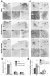
Retrograde transgene expression via HiRet and NeuRet vectors after vector injections into M2 in rats. (a) GFP immunostaining in the cortex (contralateral M2). Right panels denote higher-power magnifications of the square areas in left panels. Scale bars, 500 μm (left) and 50 μm (right). (b) Density (cells per mm2) of GFP-positive cells in the contralateral M2. Mean ± SEM (n = 4). * p < 0.05, significant differences from the value for the HiRet vector (Student’s t test). (c) GFP immunostaining in the ipsilateral thalamus (left, VL; right, CL). Right panels denote higher-power magnifications of the square areas in left panels. HC, hippocampus; R, reticular nucleus of the thalamus; 3V, third ventricle. Scale bars, 500 μm (left) and 50 μm (right). (d) Number (cells per section) of GFP-positive cells in the ipsilateral VL and CL. Data are expressed as the mean ± SEM (n = 4).
Figure 2
Figure 2
Retrograde transgene expression via HiRet and NeuRet vectors after vector injections into A6m in marmosets. (a) GFP immunostaining in the cortex (left, contralateral A6m; right, ipsilateral PE and PCC). A6DC, dorsocaudal part of area 6; A24, area 24. Scale bars, 500 µm (left) and 100 μm (right). (b) Density (cells per mm2) of GFP-positive cells in contralateral A6m and the ipsilateral PE and PCC. Data obtained in two animals are indicated with hollow and solid symbols (circles, triangles, and squares). (c) GFP immunostaining in the ipsilateral thalamus (left, VL; right, CL). Cd, caudate nucleus; LGN, lateral geniculate nucleus of the thalamus; MD, mediodorsal nucleus of the thalamus; VP, ventroposterior nucleus of the thalamus. Other abbreviations are as in Figure 1. Scale bars, 500 µm (left) and 100 μm (right). (d) Number (cells per section) of GFP-positive cells in the ipsilateral VL and CL. Data obtained in two animals are indicated with hollow and solid symbols (circles, triangles, and squares). All other conventions are as in Figure 1.
Figure 3
Figure 3
Retrograde transgene expression via HiRet and NeuRet vectors after vector injections into SMA in macaques. (a) GFP immunostaining in the cortex (ipsilateral ventral premotor cortex, PMv). Cd, caudate nucleus; PMd, dorsal premotor cortex; Put, putamen; sAS, spur of the arcuate sulcus. Scale bars, 1 mm (left) and 100 μm (right). (b) Density (cells per mm2) of GFP-positive cells in the contralateral SMA and the ipsilateral PMv. (c) GFP immunostaining in the ipsilateral thalamus (VL). All abbreviations are as in Figure 1 and Figure 2. (d) Number (cells per section) of GFP-positive cells in the ipsilateral VL and CL. Scale bars, 1 mm (left) and 100 μm (right). All other conventions are as in Figure 1.
Figure 4
Figure 4
Inflammatory/immune responses of HiRet and NeuRet vectors. (a) GFP (green) and double immunofluorescence staining for Iba1 (red) and CD8 (blue) at the injection sites of the HiRet (upper) and NeuRet (FuG-E; lower) vectors in rats. Insets: higher-power magnifications of the square areas. Scale bars: 500 μm and 100 μm for insets. (b) Data obtained for marmosets. Scale bars: 500 μm and 200 μm for insets. (c) Data obtained for macaques. HiRet vector (top), NeuRet vector (middle), and higher-titer NeuRet vector (bottom). Scale bars: 2 mm and 0.2 mm for insets.
Figure 4
Figure 4
Inflammatory/immune responses of HiRet and NeuRet vectors. (a) GFP (green) and double immunofluorescence staining for Iba1 (red) and CD8 (blue) at the injection sites of the HiRet (upper) and NeuRet (FuG-E; lower) vectors in rats. Insets: higher-power magnifications of the square areas. Scale bars: 500 μm and 100 μm for insets. (b) Data obtained for marmosets. Scale bars: 500 μm and 200 μm for insets. (c) Data obtained for macaques. HiRet vector (top), NeuRet vector (middle), and higher-titer NeuRet vector (bottom). Scale bars: 2 mm and 0.2 mm for insets.
Figure 5
Figure 5
Neuron specificity of HiRet and NeuRet vectors. (a) Double immunofluorescence histochemistry for GFP and NeuN or GFP and GFAP at the injection sites of the HiRet (upper) and NeuRet (FuG-E; lower) vectors in marmosets. Scale bar, 50 μm. (b) Ratios of double-labeled cells (GFP+NeuN+, GFP+GFAP+) to the total GFP-positive cells (GFP+). (c,d) Data obtained for macaques. Scale bar, 50 μm.

Similar articles

Cited by

References

    1. Blömer U., Naldini L., Kafri T., Trono D., Verma I.M., Gage F.H. Highly Efficient and Sustained Gene Transfer in Adult Neurons with a Lentivirus Vector. J. Virol. 1997;71:6641–6649. doi: 10.1128/jvi.71.9.6641-6649.1997. - DOI - PMC - PubMed
    1. Cockrell A.S., Kafri T. Gene Delivery by Lentivirus Vectors. Mol. Biotechnol. 2007;36:184–204. doi: 10.1007/s12033-007-0010-8. - DOI - PubMed
    1. Naldini L., Blömer U., Gage F.H., Trono D., Verma I.M. Efficient Transfer, Integration, and Sustained Long-Term Expression of the Transgene in Adult Rat Brains Injected with a Lentiviral Vector. Proc. Natl. Acad. Sci. USA. 1996;93:11382–11388. doi: 10.1073/pnas.93.21.11382. - DOI - PMC - PubMed
    1. Kobayashi K., Kato S., Inoue K., Takada M., Kobayashi K. Altering Entry Site Preference of Lentiviral Vectors into Neuronal Cells by Pseudotyping with Envelope Glycoproteins. Springer; Berlin/Heidelberg, Germany: 2016. pp. 175–186. Methods in Molecular Biology. - DOI - PubMed
    1. Mazarakis N.D., Azzouz M., Rohll J.B., Ellard F.M., Wilkes F.J., Olsen A.L., Carter E.E., Barber R.D., Baban D.F., Kingsman S.M., et al. Rabies Virus Glycoprotein Pseudotyping of Lentiviral Vectors Enables Retrograde Axonal Transport and Access to the Nervous System after Peripheral Delivery. Hum. Mol. Genet. 2001;10:2109–2121. doi: 10.1093/hmg/10.19.2109. - DOI - PubMed

Publication types

Substances

LinkOut - more resources