Review
. 2021 Jul 20;13(7):1405.
doi: 10.3390/v13071405.

Quaternary Interaction of the HIV-1 Envelope Trimer with CD4 and Neutralizing Antibodies

Affiliations
Review

Quaternary Interaction of the HIV-1 Envelope Trimer with CD4 and Neutralizing Antibodies

Qingbo Liu et al. Viruses. .

Abstract

The entry of HIV-1 into host cells is initiated by the interaction of the viral envelope (Env) spike with the CD4 receptor. During this process, the spike undergoes a series of conformational changes that eventually lead to the exposure of the fusion peptide located at the N-terminus of the transmembrane glycoprotein, gp41. Recent structural and functional studies have provided important insights into the interaction of Env with CD4 at various stages. However, a fine elucidation of the earliest events of CD4 contact and its immediate effect on the Env conformation remains a challenge for investigation. Here, we summarize the discovery of the quaternary nature of the CD4-binding site in the HIV-1 Env and the role of quaternary contact in the functional interaction with the CD4 receptor. We propose two models for this initial contact based on the current knowledge and discuss how a better understanding of the quaternary interaction may lead to improved immunogens and antibodies targeting the CD4-binding site.

Keywords: CD4; HIV-1; neutralizing antibodies; quaternary interaction; receptor binding; viral entry.


Conflict of interest statement

The authors declare no conflict of interest.

Figures
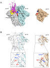
Figure 1
Figure 1
Structure of the CD4-BS2 in a SOSIP.664 Env trimer (PDB ID: 4TVP) and in a gp120 monomer (PDB ID: 4OLU). (A) Left, primary CD4-BS (yellow) and CD4-BS2 (red and blue) are shown on the trimeric Env. The CD4 molecule is docked onto the trimer by aligning its complexed gp120 moiety to one gp120 protomer of the Env trimer. Right, the amino acid residues that form CD4-BS2 are highlighted in the monomeric gp120 structure. (B) Residues of CD4-BS2 are shown as sticks and colored by their charges (blu = positive; red = negative). As shown, the side chains of key CD4-BS2 residues adopt opposite orientations in the two conformations of the gp120 subunit: outward and fully solvated in the trimer (left); inwards and predominantly solvent inaccessible in the monomer (right).
Figure 2
Figure 2
Structures of CD4 complexed with SOSIP.664 trimers in closed (5U1F), partially open or open states (6CM3, 6U0L, 5VN3, 6OPO). CD4 is colored in magenta. The three gp120 protomers of the Env trimers are colored in gray, cyan and blue, respectively. Other stabilizing ligands in each structure were removed for clarity.
Figure 3
Figure 3
Models of CD4-BS2 function in the HIV-1 entry process. (A) Hold-and-position model. Left panel: top view of a predicted unstable state with a single CD4 molecule (taken from structure 3JWD) bound only to the primary CD4-BS of the HIV-1 Env trimer (PDB ID: 4TVP). The red arrow denotes the direction of CD4 movement towards the neighboring gp120 protomer. Right panel: top view of the CD4 molecule bound with an optimal angle that engages both the primary CD4-BS and CD4-BS2. Residues on the the surface of CD4-BS2 are colored in blue and red. (B) Touch-and-go switch model: side view of a closed Env trimer (PDB ID: 4TVP) and a partially open trimer (PDB ID: 5VN3). Left panel: a monomeric gp120 structure complexed with CD4 is aligned to one protomer of the SOSIP Env trimer, showing the close contact of CD4 (yellow) with the CD4-BS2 (positively charged E62 and E64 in red, negatively charged H66 and K207 in blue). The tryptophan cavity is colored in magenta and gp41 in orange. Right panel: a partially open trimer complexed with CD4 and antibody 17b (removed for clarity). The residues are color coded as in the left panel.
Figure 4
Figure 4
Quaternary contact of anti-CD4-BS antibodies with the Env trimer. (A) Structure of VRC03 (PDB ID: 6NF2) and VRC01 (PDB ID: 5FYK) complexed with SOSIP.664 trimers. The long FR3 loop of VRC03 is colored in red. (B) Structure of FR3-03 chimeric antibodies complexed with SOSIP.664 trimers (PDB IDs: 6NM6 and 6NNF). The engrafted FR3-03 loop is shown in red.

Similar articles

Cited by

References

    1. Wilen C., Tilton J.C., Doms R.W. HIV: Cell Binding and Entry. Cold Spring Harb. Perspect. Med. 2012;2:a006866. doi: 10.1101/cshperspect.a006866. - DOI - PMC - PubMed
    1. Chen B. Molecular Mechanism of HIV-1 Entry. Trends Microbiol. 2019;27:878–891. doi: 10.1016/j.tim.2019.06.002. - DOI - PMC - PubMed
    1. Julien J.P., Cupo A., Sok D., Stanfield R.L., Lyumkis D., Deller M.C., Klasse P.J., Burton D.R., Sanders R.W., Moore J.P., et al. Crystal structure of a soluble cleaved HIV-1 envelope trimer. Science. 2013;342:1477–1483. doi: 10.1126/science.1245625. - DOI - PMC - PubMed
    1. Lyumkis D., Julien J.P., de Val N., Cupo A., Potter C.S., Klasse P.J., Burton D.R., Sanders R.W., Moore J.P., Carragher B., et al. Cryo-EM structure of a fully glycosylated soluble cleaved HIV-1 envelope trimer. Science. 2013;342:1484–1490. doi: 10.1126/science.1245627. - DOI - PMC - PubMed
    1. Pancera M., Zhou T., Druz A., Georgiev I.S., Soto C., Gorman J., Huang J., Acharya P., Chuang G.Y., Ofek G., et al. Structure and immune recognition of trimeric pre-fusion HIV-1 Env. Nature. 2014;514:455–461. doi: 10.1038/nature13808. - DOI - PMC - PubMed

Publication types

MeSH terms