. 2021 Aug 17;118(33):e2102675118.
doi: 10.1073/pnas.2102675118.

Efficient integration of transmembrane domains depends on the folding properties of the upstream sequences

Affiliations

Efficient integration of transmembrane domains depends on the folding properties of the upstream sequences

Marco Janoschke et al. Proc Natl Acad Sci U S A. .

Abstract

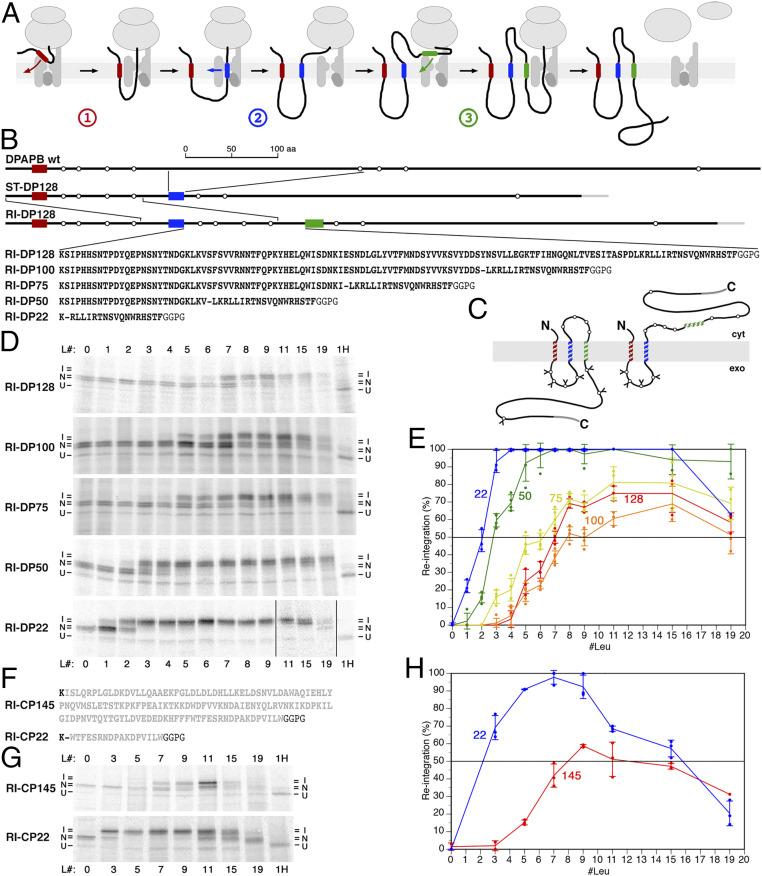
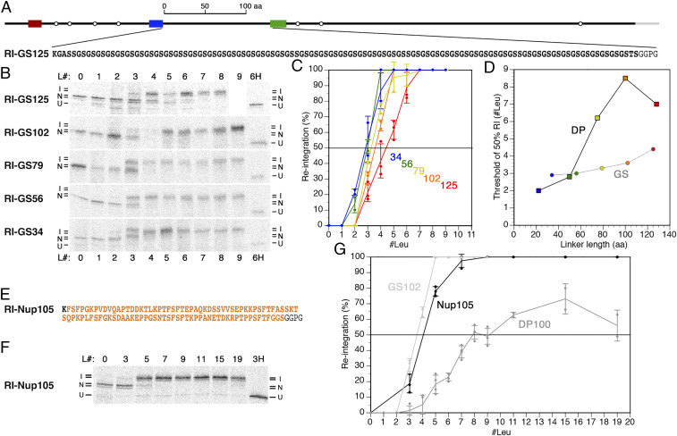
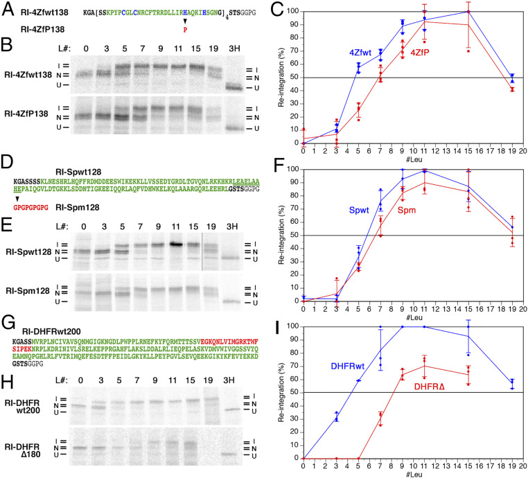
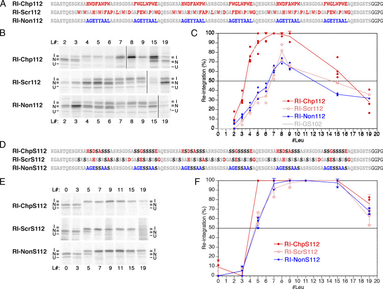
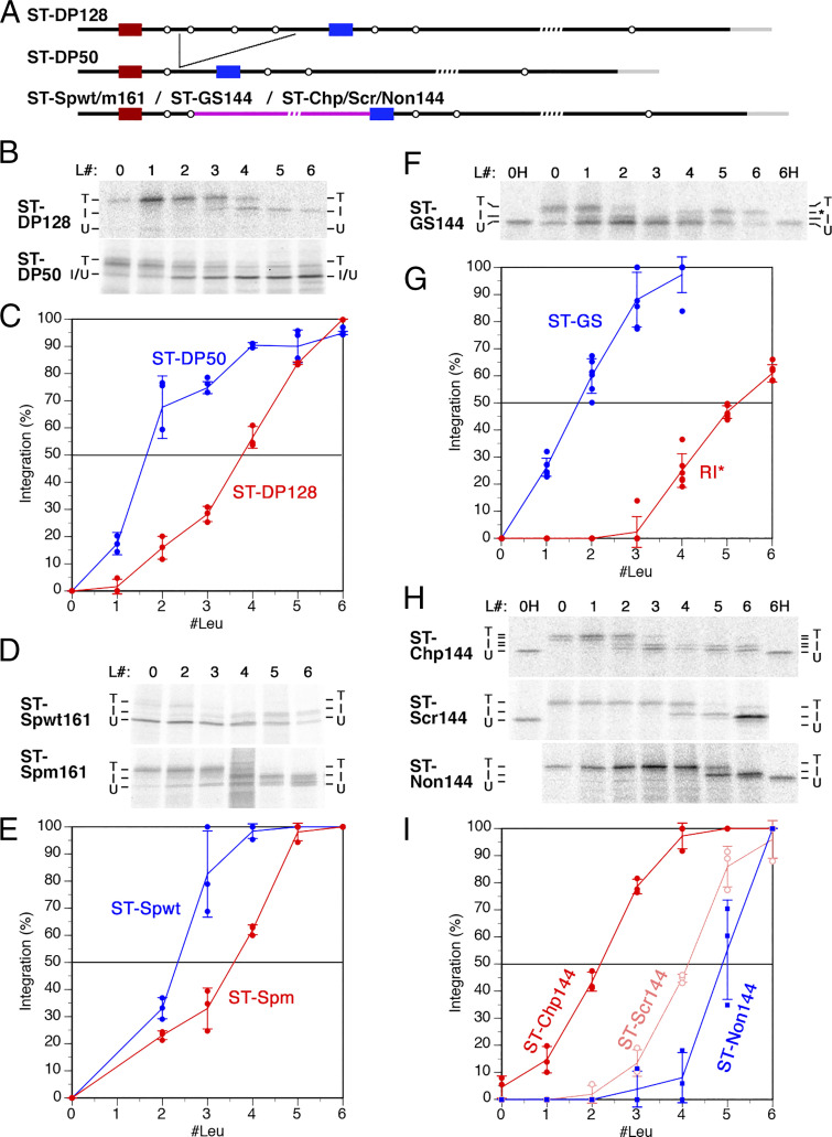
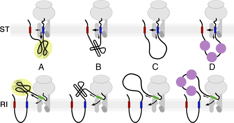
The topology of most membrane proteins is defined by the successive integration of α-helical transmembrane domains at the Sec61 translocon. The translocon provides a pore for the transfer of polypeptide segments across the membrane while giving them lateral access to the lipid. For each polypeptide segment of ∼20 residues, the combined hydrophobicities of its constituent amino acids were previously shown to define the extent of membrane integration. Here, we discovered that different sequences preceding a potential transmembrane domain substantially affect its hydrophobicity requirement for integration. Rapidly folding domains, sequences that are intrinsically disordered or very short or capable of binding chaperones with high affinity, allow for efficient transmembrane integration with low-hydrophobicity thresholds for both orientations in the membrane. In contrast, long protein fragments, folding-deficient mutant domains, and artificial sequences not binding chaperones interfered with membrane integration, requiring higher hydrophobicity. We propose that the latter sequences, as they compact on their hydrophobic residues, partially folded but unable to reach a native state, expose hydrophobic surfaces that compete with the translocon for the emerging transmembrane segment, reducing integration efficiency. The results suggest that rapid folding or strong chaperone binding is required for efficient transmembrane integration.

Keywords: Sec61 translocon; membrane proteins; molecular chaperones; protein folding; topogenesis.


Conflict of interest statement

The authors declare no competing interest.

Figures

Fig. 1.
Fig. 1.
Reintegration efficiency is affected by the cytoplasmic upstream sequence. (A) Schematic representation of the three distinct integration processes in the biogenesis of membrane proteins: signal (anchor) integration (red transmembrane segment) (1), stop-transfer integration (blue) (2), and reintegration (green) (3). Transmembrane domains do not necessarily fully enter the translocon, before exiting into the membrane, but may associate with lipids early on and glide along the gate, as proposed by Cymer et al. (43). (B) The construct RI-DP128 was derived from wt DPAPB via ST-DP128 (DPAPB-H in ref. 12), as shown schematically with the transmembrane domains in red, blue, and green as in A. Rings indicate potential N-glycosylation sites. A C-terminal triple–HA-tag is shown in gray. To analyze the hydrophobicity threshold of membrane integration, the H segment sequence GGPGAAAAAAAAAAAAAAAAAAAGPGG, with 0 to 19 alanines replaced by leucines, were inserted as a potential reintegration sequence (green) in RI-DP. Below, the DPAPB sequences of the cytosolic loop between the stop-transfer and the reintegration domains of RI-DP128 and the deletion constructs RI-DP100–22 are listed, with a dash indicating the deletion site. At least 20 residues preceding the reintegration H segment were kept constant in all constructs. (C) Schematic representation of the two topologies when the H segments do or do not initiate reintegration and C-terminal translocation. cyt, cytosol; exo, exoplasm = ER lumen. Glycosylations are indicated by Y. (D) The various constructs, each with different reintegration H segments composed of 19 alanines, with 0 to 19 of them replaced by leucine residues (L#), were expressed in yeast cells, labeled with [35S]methionine for 5 min, immunoprecipitated, and analyzed by SDS–gel electrophoresis and autoradiography. Based on the glycosylation pattern, integration (I) or nonintegration (N) of the H segment, as well as uninserted unglycosylated products (U) were distinguished. To identify the position of the unglycosylated polypeptide, a sample was analyzed after deglycosylation by endoglycosidase H (H) on the right. (E) The fraction of products with reintegrated H segments as a percentage of the total membrane-integrated proteins was plotted versus the number of leucine residues in the H segment (mean, SD, and the individual values of at least three independent experiments). The curves are labeled with the length of the constructs’ cytosolic loops between stop transfer and H segment. (F) The cytoplasmic sequence between stop transfer and H segment is shown for RI-CP145 (residues 21 to 160 of pre-CPY, in gray) and RI-CP22 (residues 144 to 160). (G) The CPY constructs were expressed and analyzed, as in D. (H) Reintegration of the H segment was quantified, as in E (mean, SD, and the individual values of at least three independent experiments).
Fig. 2.
Fig. 2.
Reintegration after a generic glycine-serine repeat or a natural intrinsically disordered sequence is efficient and largely length independent. (A) The construct RI-GS125 is schematically illustrated, and its generic glycine-serine repeat sequence between stop transfer and H segment is shown. (B) RI-GS125 and constructs with truncated GS repeat sequences (RI-GS102–34) were expressed, radioactively labeled, and analyzed, as in Fig. 1D. L# indicates the number of leucines in the H segment. Based on the glycosylation pattern, integration (I) or nonintegration (N) of the H segment, as well as uninserted unglycosylated products (U) were distinguished. Construct RI-GS125-L9 was not obtained, and the corresponding lane was thus replaced by white space. (C) The fraction of H segment reintegration was plotted versus the number of leucine residues in the H segment (mean, SD, and the individual values of at least three independent experiments). (D) The interpolated hydrophobicity threshold for 50% reintegration is plotted versus the linker length between stop transfer and H segment for the DP constructs (squares; from Fig. 1E) and the GS constructs (from C). (E) The sequence between stop transfer and H segment, containing residues 614 to 711 of Nup1p. (F) Autoradiographs of RI-Nup105 constructs with different H segments after radioactive labeling and gel electrophoresis. (G) The fraction of H segment reintegration was plotted versus the number of leucine residues in the H segment (mean, SD, and the individual values of at least three independent measurements).
Fig. 3.
Fig. 3.
Effect of rapidly folding or mutated domains on the reintegration of a subsequent H segment. (A) Four consecutive copies of a zinc finger domain of yeast Adr1p (shown in green with the Zn2+-coordinating residues in blue) were inserted between the stop-transfer sequence and the reintegration H segment in construct RI-4Zfwt138. In RI-4ZfP138, one Zn2+-coordinating histidine was mutated to proline (in red) in each zinc finger to prevent folding. (B) Autoradiographs of the wt and mutant zinc finger constructs with different H segments are shown, as in Fig. 1D. L# indicates the number of leucines in the H segment. Based on the glycosylation pattern, integration (I) or nonintegration (N) of the H segment, as well as uninserted unglycosylated products (U) were distinguished. (C) The fraction of H segment reintegration was plotted versus the number of leucine residues in the H segment (mean, SD, and the individual values of at least three independent experiments). (D–F) Spectrin domain 16 of human spectrin α-chain (green) was inserted between the stop-transfer sequence and the reintegration H segment in construct RI-Spwt128. For RI-Spm128, nine underlined, central residues were replaced by a glycine/proline repeat sequence (red) to prevent native folding. Autoradiographs of the expressed proteins with different H segments were analyzed and the fraction of H segment reintegration quantified as above (B and C). (G–I) The wt DHFR was inserted between the stop-transfer sequence and the reintegration H segment in construct RI-DHFRwt200. In RI-DHFR∆180, the red sequence was deleted. Autoradiographs of the expressed proteins with different H segments were analyzed and the fraction of H segment reintegration quantified as above (B and C).
Fig. 4.
Fig. 4.
Chaperone-binding sequences facilitate reintegration. (A) Three repeats of two different octapeptides (red) that have been shown to bind with high affinity to BiP (29), separated by hydrophilic octapeptides (gray), were inserted between the stop-transfer sequence and the reintegration H segment in construct RI-Chp112. In RI-Scr112, the sequence was scrambled to abolish chaperone binding. In RI-Non112, repeats of an octapeptide were used that had been shown not to bind BiP (blue) (29). (B) Autoradiographs of the above constructs with different H segments are shown, as in Fig. 1D. L# indicates the number of leucines in the H segment. Based on the glycosylation pattern, integration (I) or nonintegration (N) of the H segment, as well as uninserted unglycosylated products (U) were distinguished. (C) The fraction of H segment reintegration was plotted versus the number of leucine residues in the H segment (mean, SD, and the individual values of at least three independent experiments). For comparison, the tracing of the curve for RI-GS102 of the similar loop length from Fig. 2C is indicated in gray. (D–F) In constructs RI-ChpS112/ScrS112/NonS112, the hydrophobic residues (L, F, P, and W) in the segment between stop-transfer sequence and reintegration H segment were replaced by serines (in black; D). Upon expression and autoradiography (E), the fraction of H segment reintegration (F) was plotted, as above.
Fig. 5.
Fig. 5.
Stop-transfer integration similarly depends on the folding properties of the upstream sequence as reintegration. (A) Schematic representation of the original stop-transfer construct ST-DP128 with a DPAPB fragment of 128 residues separating the signal anchor (red) from the stop-transfer H segment (blue). In ST-DP50, the length of this spacer was truncated to 50 residues as indicated. In addition, constructs with a wt or a mutant spectrin domain (ST-SPwt161 and ST-Spm161, respectively; Fig. 3D), with a 100-residue glycine-serine repeat sequence (ST-GS144; Fig. 2A) and the chaperone-binding, -scrambled, or -nonbinding sequences (ST-Chp/Scr/Non144; Fig. 4A), were inserted into the luminal spacer after the second glycosylation site (pink line). The number in the names indicates the total lengths between stop transfer and H segment. (B–I) The indicated constructs were expressed, labeled with [35S]methionine, and analyzed by SDS–gel electrophoresis and autography (B, D, F, and H). L# indicates the number of leucines in the H segment. Based on the glycosylation pattern, integration (I) or translocation (T) of the H segment, as well as uninserted unglycosylated products (U) were distinguished. The fraction of H segment integration was plotted versus the number of leucine residues in the H segment (mean, SD, and the individual values of at least three independent experiments; C, E, G, and I). Stop-transfer integration was calculated as the twice-glycosylated (loop translocated) products, as a percentage of both loop-translocated and fully translocated products. For ST-DP50 (B), the single potential glycosylation site in the spacer was found not functional. Stop-transfer integration thus resulted in an increase of the unglycosylated fraction, which was thus used as a measure of integration. For ST-GS144 (F), an additional product with three glycans (*) was increasingly obtained with H segments containing ≥4 leucines. This form most likely corresponds to products that failed to translocate the luminal loop but instead translocated the C-terminal domain, as a result of the H segment acting as a reintegration sequence. Stop-transfer efficiency was determined as the fraction of the twice-glycosylated (loop translocated) products of the sum of proteins with two and five glycans, while reintegration (RI*; shown in gray) was determined as the percentage of threefold glycosylated products of the sum of proteins with three and zero glycans (G).
Fig. 6.
Fig. 6.
How the folding state of upstream sequences affects membrane integration. A nascent chain emerging from the ribosome (in the case of reintegration; RI) or from the luminal side of the translocon (in the case of stop transfer; ST) will compact vectorially, acquire some secondary and tertiary structure but remain conformationally fluctuating, and expose substantial hydrophobic surfaces that—if the sequence cannot reach a native fold (as for protein fragments or mutant domains)—will compete with translocon/membrane for the transmembrane segment and affect integration efficiency (A). If the sequence is folded rapidly (B), if it is intrinsically disordered (C), or if the sequence is bound by chaperones (D), they interact little with potential transmembrane domains, thus allowing efficient integration with low-hydrophobicity thresholds.

Comment in

References

    1. Spiess M., Junne T., Janoschke M., Membrane protein integration and topogenesis at the ER. Protein J. 38, 306–316 (2019). - PubMed
    1. Rapoport T. A., Li L., Park E., Structural and mechanistic insights into protein translocation. Annu. Rev. Cell Dev. Biol. 33, 369–390 (2017). - PubMed
    1. Voorhees R. M., Fernández I. S., Scheres S. H. W., Hegde R. S., Structure of the mammalian ribosome-Sec61 complex to 3.4 Å resolution. Cell 157, 1632–1643 (2014). - PMC - PubMed
    1. Gogala M., et al. ., Structures of the Sec61 complex engaged in nascent peptide translocation or membrane insertion. Nature 506, 107–110 (2014). - PubMed
    1. Voorhees R. M., Hegde R. S., Structure of the Sec61 channel opened by a signal sequence. Science 351, 88–91 (2016). - PMC - PubMed

Publication types

Substances

LinkOut - more resources