Review
. 2021;46(3):79.
doi: 10.1007/s12038-021-00204-z.

Connecting primitive phase separation to biotechnology, synthetic biology, and engineering

Affiliations
Review

Connecting primitive phase separation to biotechnology, synthetic biology, and engineering

Tony Z Jia et al. J Biosci. 2021.

Abstract

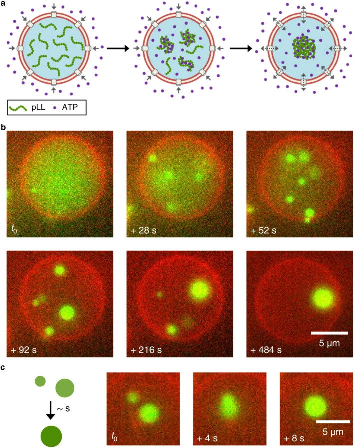
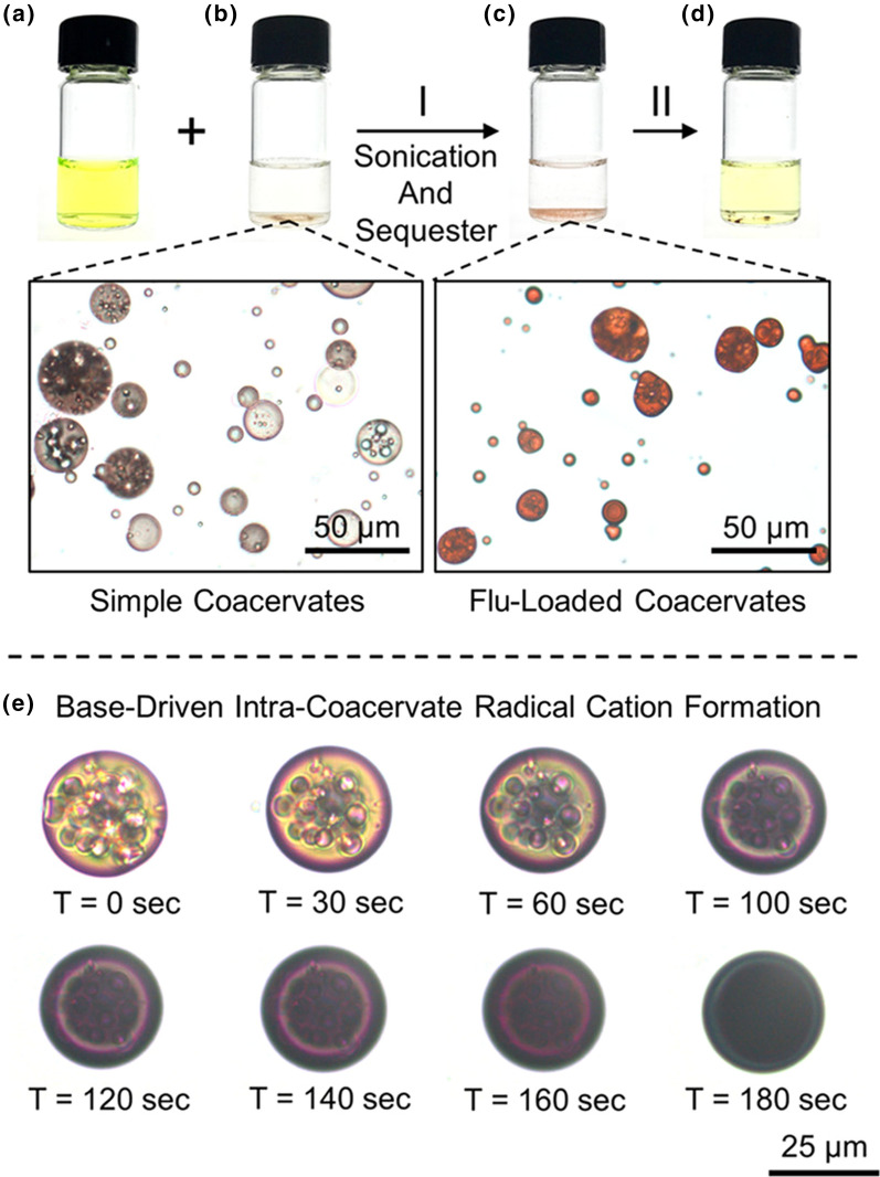
One aspect of the study of the origins of life focuses on how primitive chemistries assembled into the first cells on Earth and how these primitive cells evolved into modern cells. Membraneless droplets generated from liquid-liquid phase separation (LLPS) are one potential primitive cell-like compartment; current research in origins of life includes study of the structure, function, and evolution of such systems. However, the goal of primitive LLPS research is not simply curiosity or striving to understand one of life's biggest unanswered questions, but also the possibility to discover functions or structures useful for application in the modern day. Many applicational fields, including biotechnology, synthetic biology, and engineering, utilize similar phaseseparated structures to accomplish specific functions afforded by LLPS. Here, we briefly review LLPS applied to primitive compartment research and then present some examples of LLPS applied to biomolecule purification, drug delivery, artificial cell construction, waste and pollution management, and flavor encapsulation. Due to a significant focus on similar functions and structures, there appears to be much for origins of life researchers to learn from those working on LLPS in applicational fields, and vice versa, and we hope that such researchers can start meaningful cross-disciplinary collaborations in the future.


Figures

Figure 1
Figure 1
Jeewanu particles after 24 hours of sunlight exposure as observed by different microscopy methods. Left: Light microscopy. Middle: Confocal microscopy. Right: Scanning Electron Microscopy (SEM). Figures reproduced with permission from Gupta VK and Rai R K 2018 Cytochemical characterisation of photochemically formed, self-sustaining, abiogenic, protocell-like, supramolecular assemblies ‘Jeewanu’. Int. J. Life Sci. 6(4):877–884 (Gupta and Rai 2018) under a Creative Commons License.
Figure 2
Figure 2
Formation of membraneless droplets through segregative (top; (a) and (b)), e.g. ATPS, and associative (bottom; (c) and (d)), e.g., complex coacervates, phase separation. In segregative phase separation, the polymers and molecules involved in the phase separation process are generally confined to separate phases (generally due to thermodynamic reasons). In associative phase separation, the polymers and molecules involved in the phase separation process generally interact and reside in the same condensed phase, with a dilute aqueous phase surrounding the concentrated droplet. Figures reproduced with permission from Crowe CD and Keating CD 2018 Liquid–liquid phase separation in artificial cells. Interface Focus 8(5):20180032 (Crowe and Keating 2018). Copyright The Royal Society 2018.
Figure 3
Figure 3
Use of amylose-based coacervates for protein therapy delivery to cells. Oppositely charged cationic Q-Amylose and anionic Cm-Amylose polymers interact (top) to form a phase-separated coacervate (bottom right). This phase coacervate can then be loaded by protein therapies, such as myoglobin, to be delivered to specific cells, such as human stem cells. Figure reproduced with permission from Xiao W et al. 2020 Biopolymeric coacervate microvectors for the delivery of functional proteins to cells. Adv. Biosyst. 4(11):2000101 (Xiao et al. 2020) under a Creative Commons License.
Figure 4
Figure 4
An enzymatic cascade taking place within a coacervate-in-ATPS system. GOx first oxidizes glucose (originally in the PEG phase) within the dextran droplet to form hydrogen peroxide (H2O2). The H2O2 then migrates into the coacervate phase (ATP-Poly(diallyldimethylammonium chloride)), and is used as an oxidating co-factor for HRP to oxidize an inactive reporter dye (Amplex Red, ABTS, oPD, or DAB) to its active fluorescent or colorometric form. Figure reproduced with permission from Kojima K and Takayama S 2018 Membraneless compartmentalization facilitates enzymatic cascade reactions and reduces substrate inhibition. ACS Appl. Mater. Interfaces 10(38):32782–32791 (Kojima and Takayama 2018). Copyright American Chemical Society 2018.
Figure 5
Figure 5
Formation of a coacervate within a liposome. (a) ATP flows into a liposome through α-hemolysin pores, interacting and complexing with poly-L-lysine (pLL), phase separating to form a coacervate within the liposome. (b) Formation of the coacervate droplets (green) within a liposome over time. (c) Coalescence of two separate coacervate droplets within a liposome over time. An enzymatic cascade taking place within a coacervate-in-ATPS system. GOx first oxidizes glucose (originally in the PEG phase) within the dextran droplet to form hydrogen peroxide (H2O2). The H2O2 then migrates into the coacervate phase (ATP-Poly(diallyldimethylammonium chloride)), and is used as an oxidating co-factor for HRP to oxidize an inactive reporter dye, Amplex Red, to its active fluorescent form (Resolufin). Figure reproduced with permission from Deshpande, S. 2019. ‘Spatiotemporal control of coacervate formation within liposomes.’ Nat. Commun. 10:1800 (Deshpande et al. 2019) under a Creative Commons License.
Figure 6
Figure 6
Sequestration of fluorescein dye within a bipyridinium-functionalized poly-lipoic ester (BPLE) coacervate through sonication. (a) Fluorescein in solution, as visible by the yellow hue. (b) BPLE coacervate droplets in solution (bottom left micrograph). (c) After combining the coacervates and fluorescein, sonication results in fluorescein becoming sequestered within the droplets (bottom right micrograph). (d) Addition of acetic acid to the fluorescein-loaded coacervates results in release of fluorescein back into solution, as visible by the off-yellow hue. (e) Coacervates exposed to pH 14 conditions results in rapid formation of radical cations within the droplets, as observed by the coacervates changing to a violet color over time. Figure reproduced with permission from Zhang Z et al. 2019 Poly-lipoic ester-based coacervates for the efficient removal of organic pollutants from water and increased point-of-use versatility. Chem. Mater. 31(12):4405–4417 (Zhang et al. 2019). Copyright American Chemical Society 2019.

Similar articles

Cited by

References

    1. Abbas M, Lipiński WP, Wang J, Spruijt E. Peptide-based coacervates as biomimetic protocells. Chem. Soc. Rev. 2021;50:3690–3705. doi: 10.1039/D0CS00307G. - DOI - PubMed
    1. Afify A, Gwad EL, AA, and EL Rahman NA, Using Enterobacter aerogenes DSM 30053 for bio-hydrogen production by microbial electrolysis cells from domestic wastewater. J. Agric. Chem. Biotech. 2017;8:167–171.
    1. Akama Y, Sali A. Extraction mechanism of Cr(VI) on the aqueous two-phase system of tetrabutylammonium bromide and (NH(4))(2)SO(4) mixture. Talanta. 2002;57:681–686. doi: 10.1016/S0039-9140(02)00076-0. - DOI - PubMed
    1. Alberts B, Johnson AD, Lewis J, Morgan D, Raff M, Roberts K, Walter P. Molecular biology of the cell: sixth international student. New York: W.W. Norton & Company; 2014.
    1. Alberts B, Johnson A, Lewis J, Raff M, Roberts K, and Walter P 2007 Molecular biology of the cell (New York: Garland Science)