. 2021 Aug 9;12(1):4795.
doi: 10.1038/s41467-021-24690-9.

A genetically encoded tool for reconstituting synthetic modulatory neurotransmission and reconnect neural circuits in vivo

Affiliations

A genetically encoded tool for reconstituting synthetic modulatory neurotransmission and reconnect neural circuits in vivo

Josh D Hawk et al. Nat Commun. .

Abstract

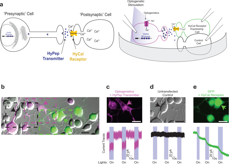
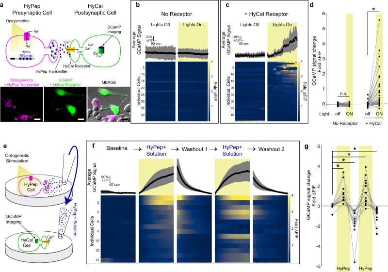
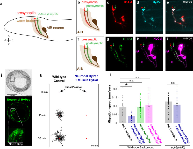
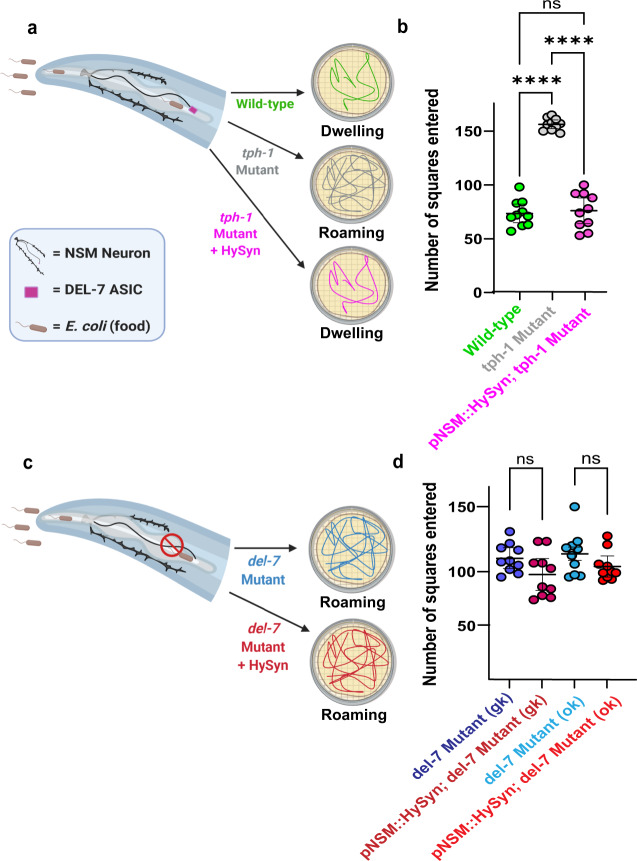
Chemogenetic and optogenetic tools have transformed the field of neuroscience by facilitating the examination and manipulation of existing circuits. Yet, the field lacks tools that enable rational rewiring of circuits via the creation or modification of synaptic relationships. Here we report the development of HySyn, a system designed to reconnect neural circuits in vivo by reconstituting synthetic modulatory neurotransmission. We demonstrate that genetically targeted expression of the two HySyn components, a Hydra-derived neuropeptide and its receptor, creates de novo neuromodulatory transmission in a mammalian neuronal tissue culture model and functionally rewires a behavioral circuit in vivo in the nematode Caenorhabditis elegans. HySyn can interface with existing optogenetic, chemogenetic and pharmacological approaches to functionally probe synaptic transmission, dissect neuropeptide signaling, or achieve targeted modulation of specific neural circuits and behaviors.


Conflict of interest statement

The authors declare no competing interests.

Figures

Fig. 1
Fig. 1. Hydra-derived synthetic synapse (HySyn) engineered through heterologous expression of Hydra neuropeptide (HyPep) and receptor (HyCal).
a (left) Schematic describing “HySyn”: Hydra-derived neuropeptide (blue) is released from the “presynaptic” cell and interacts with the HyCal receptor (yellow) to flux calcium in the “postsynaptic” cell. a (right) Schematic illustrating the experimental paradigm to assay HySyn function by whole-cell patch-clamp electrophysiology. b Micrograph showing co-culture of Neuro2a neuroblastoma cells expressing either “presynaptic” HyPep with ChRoME (magenta), “postsynaptic” HyCal with GFP (green), or untransfected (grayscale). Boxed regions correspond to the micrographs in ce, with the color of the box matched to the color of the title of the micrograph. ce Identification of cell populations based on fluorescent markers (top, micrograph, 20μm scale bar) enabling whole-cell current recording (bottom, “current traces”) from three distinct populations during optogenetic stimulation (480 nm, blue-shaded windows in “current traces”, “On”): c “presynaptic” cells expressing HyPep with ChRoME (pseudocolored magenta) consistently produced step-wise optogenetic currents, d unlabeled cells remain unaltered by optogenetic activation of neighboring HyPep+ cells, and e green “postsynaptic” cells expressing the HyCal receptor show an increasing inward current during light stimulation. The downward deflection of traces during optogenetic stimulation in blue windows indicates an inward current, suggesting a depolarizing cation current. The persistence of HyCal current between stimulation (e) is consistent with the lack of desensitization of HyCal channel and suggests an accumulation of HyPep neuropeptide in the bath solution. Transfection and reporter expression (micrographs in be, see also Fig. 2a) was reproducibly observed, including in the 22 cases where successful electrophysiological recordings were made (Supplementary Fig. 2), and in 3 independent populations with GCaMP expression (Fig. 2).
Fig. 2
Fig. 2. HySyn produces volumetric neuromodulation of postsynaptic calcium.
a Schematic illustrating the experimental paradigm used to characterize HyPep to HyCal signaling by using calcium imaging, top. Micrograph illustrating transfected Neuro2a cells used in these experiments, bottom (20 μm scale bar). Chrimson was used for optogenetic stimulation (591 nm, 500 ms at 1 Hz) of “presynaptic” cells expressing the HyPep carrier of the neuropeptide (“Hydra RFamide”). GCaMP was expressed in distinct cells either alone (b) or with the cognate receptor HyCal (c), and both of these two groups of cells were co-cultivated with HyPep “presynaptic” cells. b Average GCaMP signals, top bold line with shaded 95% confidence intervals, and individual cell heatmap profiles, bottom, for Neuro2a cells expressing GCaMP alone. Without receptor expression, but even in the presence of HyPep-secreting cells co-expressing the optogenetic tool (Chrimson), GCaMP signals remained stable in these cells both without (left, “Lights Off”) and with (right, “Lights On”, yellow shading) optogenetic stimulation of the HyPep cells. c As in b, but in cells also expressing the HyCal receptor in the presence of HyPep-secreting cells co-expressing the optogenetic tool (Chrimson). We observe calcium signal rise over the course of light stimulation. Out of 44 cells, ~34% (14) showed changes in fluorescence over 3 standard deviations beyond the mean change observed prior to light stimulation. d Quantification of the change in GCaMP signal (mean ΔF/F in final 30 s minus mean ΔF/F in first 30 s) revealed that optogenetic stimulation of co-cultured HyPep-expressing cells co-expressing the optogenetic tool (Chrimson) did not result in stimulation of potential “postsynaptic” cells lacking the HyCal receptor (but expressing GCaMP). In contrast, they resulted in a doubling of intracellular calcium signal in cells expressing the HyCal receptor (p = 1.0 for “No Receptor” (n = 43) Lights off vs ON, p = 0.00002 for “+HyCal Receptor” (n = 44) Lights off vs ON). e Schematic illustration of solution transfer experiments. After optogenetic stimulation (as in a for 5 min), the bathing solution from HyPep-expressing cells (“HyPep+ Solution”) was transferred to naive “postsynaptic” cells in another culture dish expressing GCaMP and the HyCal receptor. f Following a stable baseline, applying the HyPep+ solution increased the GCaMP signal, and this rise was reversed after washout and applying the fresh bathing solution (Washout). Repeated cycles of washout and application of the HyPep+ solution reproducibly increased the GCaMP signal in the same responding cells (n = 14, p = 0.00004 1st addition, p = 0.0001 Washout 1, p = 0.0003 2nd addition, p = 0.0003 Washout 2). g To highlight the kinetics of individual responding cells (from panel f), we quantified (as in d) GCaMP changes for cells that displayed responses. Error bars (and shaded regions in b, c, f) indicate 95% confidence intervals and * indicates p < 0.05 using two-tailed Mann–Whitney–Wilcoxon test with Bonferroni correction for multiple comparisons. Transfection and reporter expression (micrographs in a) were reproducibly observed, including in the 22 cases where successful electrophysiological recordings were made (Supplementary Fig. 2), and in 3 independent populations with GCaMP expression (Fig. 2). Source data are provided as a Source Data file.
Fig. 3
Fig. 3. HySyn components localize in vivo to appropriate synaptic compartments, and modify animal behavior.
a Schematic of the worm head (black outline) illustrating the polarized distribution of synaptic specializations in the neuron AIB (brown) as determined by previous cell biological and electron microscopy studies,. Presynaptic sites are restricted to the region of the neurite most distal to the soma (indicated in red, “presynaptic”). Postsynaptic specializations are enriched in the posterior soma-proximal region of the neurite (indicated in green, “postsynaptic”). be Schematic illustrating enrichment of presynaptic specializations in the neuron AIB (b red shading) and representative confocal images of the presynaptic dense-core vesicle marker IDA-1/phogrin::mCherry, (c red), and the presynaptic component of HySyn, HyPep-eGFP (d cyan). Overlay (e “merge”) showing colocalization of these markers in the presynaptic compartment of the neurite (red arrow marks the position of the distal neurite enriched for presynaptic specializations, as shown in schematic in b). The scale bar in c is 10 µm. fi Schematic illustrating enrichment of postsynaptic specializations in the neuron AIB (f green shading) and representative confocal images of the postsynaptic receptor GLR-1::GFP (g green), and the postsynaptic component of HySyn, HyCal-mCherry (h magenta). Overlay (i merge) showing that within the AIB neurite these two elements colocalize predominantly to the soma-proximal postsynaptic region (green arrow marks the position of the proximal neurite enriched for postsynaptic specializations, as shown in the schematic in f). Bright-field (j top) and fluorescence (j bottom) micrographs show the pattern of expression for HyPep-GFP under the control of a pan-neuronal promoter in the nematode C. elegans. The fluorescence pattern (green, bottom panel) shows a dense collection of puncta in the nerve ring (inset), a synaptic-rich neuropil. k Illustration of trajectories for 24 worms over a 30 min observation period. Each line represents a single worm track from a commonly aligned initial position (red dot) after either a 0 min (top), 15 min (middle), or 30 min (bottom) monitoring period. Compared to the dispersion of wild-type control (left), transgenic animals expressing the synthetic HySyn connection between neurons and muscles (right, “Neuronal HyPep + Muscle HyCal”) showed substantially reduced migration over time. l During infrequent bouts of detectable migration in this 30 min interval, those animals expressing the full HySyn system (“Neuronal HyPep + Muscle HyCal”, blue) move at a slower speed than control animals without HySyn (“No Transgene”, black) (p = 0.000001 for wild-type (n = 43) vs “Neuronal HyPep + Muscle HyCal” (n = 24)). Neither the neuropeptide itself (“Neuronal HyPep”, green) nor the receptor in the presence of intestine-produced neuropeptide (“Intestinal HyPep + Muscle HyCal”, magenta) altered migration speeds (p = 1.0 for wild-type (n = 43) vs “Neuronal HyPep” (n = 68), p = 1.0 for wild-type (n = 43) vs “Intestinal HyPep + Muscle HyCal” (n = 45)). Migration in animals carrying a mutation of egl-3(n150), a gene required for neuropeptide maturation. The egl-3(n150) mutation suppressed the function of the reconstituted HyPep-HyCal connection (right-most bar, blue) based on comparison of egl-3(n150) mutants with “Neuronal HyPep + Muscle HyCal” (n = 37) to wild-type (n = 43, p = 1.0) or egl-3(n150) mutant animals without transgene expression (n = 52, p = 0.1243). Error bars indicate 95% confidence intervals and * indicates p < 0.05 using two-tailed Mann–Whitney–Wilcoxon test with Bonferroni correction for multiple planned comparisons. Source data are provided as a Source Data file.
Fig. 4
Fig. 4. Suppression of abnormal behavioral states via targeted reconstitution of HySyn neuromodulatory connections.
a A schematic of a nematode head, and the pharyngeal, food-sensing enteric neuron NSM (black). NSM senses the presence of bacteria (food) via the DEL-7 acid-sensing ion channel (ASIC) receptor (purple box), and releases serotonin, resulting in a “dwelling” behavioral state in wild-type animals (green), which can be scored by quantifying animal displacement in the food-covered Petri dishes, as depicted in the schematic and described,. Mutant animals for serotonin biosynthesis, such as tph-1(mg280), are incapable of releasing serotonin and remain in a state of “roaming” even in the presence of food (gray). Reconstitution of circuit connectivity by using HySyn to connect serotonin-releasing NSM neuron and muscles. Note this configuration of HySyn can suppress the abnormal “roaming” state in tph-1(mg280) mutants. b Quantification of the roaming and dwelling phenotypes, as described,, for the indicated genotypes. Each dot in the graph represents an individual animal, and a total of ten animals were blindly scored per genotype (p < 0.0001 for tph-1 mutant vs. wild-type, p < 0.0001 for tph-1 mutant vs. pNSM::HySyn; tph-1 mutant, n = 10 independent biological replicates for all groups). c As a, but for mutant animals lacking the del-7 receptor, which makes them incapable of sensing food. Note that these animals are wild-type for the serotonin biosynthetic pathway, but phenocopy serotonin mutant animals because the NSM enteric neuron is not activated in the presence of food,. d As b, but for two comparable alleles of del-7 (n = 10 independent biological replicates for all groups). del-7(ok1187) (light blue) contains a deletion for the intercellular region of DEL-7, and del-7(gk688559) (dark blue) contains an early stop codon. Note that del-7 mutants display roaming behaviors, but that the reconstituted HySyn (dark and light red), as expected, is incapable of suppressing del-7, as del-7 mutant animals are incapable of sensing food, incapable of activating NSM and therefore incapable of inducing the release of HyPep in the presence of food. This experiment demonstrates that the HySyn suppression of the tph-1 mutants results from the activity-dependent release of HyPep from NSM upon encountering food. One-way ANOVA followed by a Tukey’s multiple comparison post hoc test was used to compare the means of each group. * indicates p < 0.05, ns indicates that no statistically significant difference was observed. Error bars (black) represent the mean of each group and 95% confidence intervals. Schematics were made with BioRender. Source data are provided as a Source Data file.

Similar articles

Cited by

References

    1. Roth BL. DREADDs for neuroscientists. Neuron. 2016;89:683–694. doi: 10.1016/j.neuron.2016.01.040. - DOI - PMC - PubMed
    1. Kim CK, Adhikari A, Deisseroth K. Integration of optogenetics with complementary methodologies in systems neuroscience. Nat. Rev. Neurosci. 2017;18:222–235. doi: 10.1038/nrn.2017.15. - DOI - PMC - PubMed
    1. Talay M, et al. Transsynaptic mapping of second-order taste neurons in flies by trans-tango. Neuron. 2017;96:783–795. doi: 10.1016/j.neuron.2017.10.011. - DOI - PMC - PubMed
    1. Macpherson LJ, et al. Dynamic labelling of neural connections in multiple colours by trans-synaptic fluorescence complementation. Nat. Commun. 2015;6:10024. doi: 10.1038/ncomms10024. - DOI - PMC - PubMed
    1. Rabinowitch I, Schafer WR. Engineering new synaptic connections in the C. elegans connectome. Worm. 2015;4:e992668. doi: 10.4161/21624054.2014.992668. - DOI - PMC - PubMed

Publication types