

Deep learning-enabled ultra-widefield retinal vessel segmentation with an automated quality-optimized angiographic phase selection tool
- PMID: 34373610
- PMCID: PMC9391395
- DOI: 10.1038/s41433-021-01661-4
Deep learning-enabled ultra-widefield retinal vessel segmentation with an automated quality-optimized angiographic phase selection tool
Abstract
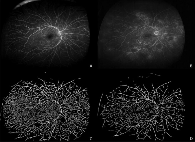
Objectives: To demonstrate the feasibility of a deep learning-based vascular segmentation tool for UWFA and evaluate its ability to automatically identify quality-optimized phase-specific images.
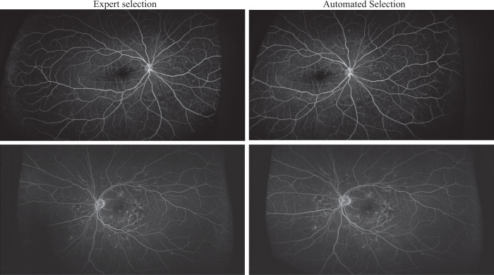
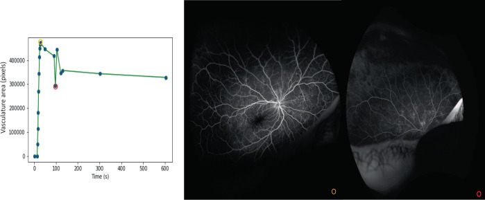
Methods: Cumulative retinal vessel areas (RVA) were extracted from all available UWFA frames. Cubic splines were fitted for serial vascular assessment throughout the angiographic phases of eyes with diabetic retinopathy (DR), sickle cell retinopathy (SCR), or normal retinal vasculature. The image with maximum RVA was selected as the optimum early phase. A late phase frame was selected at a minimum of 4 min that most closely mirrored the RVA from the selected early image. Trained image analysts evaluated the selected pairs.
Results: A total of 13,980 UWFA sequences from 462 sessions were used to evaluate the performance and 1578 UWFA sequences from 66 sessions were used to create cubic splines. Maximum RVA was detected at a mean of 41 ± 15, 47 ± 27, 38 ± 8 s for DR, SCR, and normals respectively. In 85.2% of the sessions, appropriate images for both phases were successfully identified. The individual success rate was 90.7% for early and 94.6% for late frames.
Conclusions: Retinal vascular characteristics are highly phased and field-of-view sensitive. Vascular parameters extracted by deep learning algorithms can be used for quality assessment of angiographic images and quality optimized phase selection. Clinical applications of a deep learning-based vascular segmentation and phase selection system might significantly improve the speed, consistency, and objectivity of UWFA evaluation.
© 2021. The Author(s), under exclusive licence to The Royal College of Ophthalmologists.
Conflict of interest statement
SKS receives funding from Gilead, Regeneron, and Allergan; receives compensation as a consultant from Bausch and Lomb and Santen; owns a patent with Leica. CW receives compensation as a consultant from Adverum, Allergan, Apellis, Clearside, EyePoint, Genentech/Roch, Neurotech, Novartis, Opthea, Regeneron, Regenxbio, Samsung, Santen, Alimera Sciences, Allegro, Alynylam, Bayer, Clearside, D.O.R.C., Kodiak, Notal Vision, ONL Therapeutics, PolyPhotonix, and RecensMedical. AWS receives compensation as a consultant from Allergan and Novartis. AV is an employee of ERT. JPE receives funding and compensation as a consultant from Aerpio, Adverum Alcon, Thrombogenics/Oxurion, Regeneron, Stealth, Roche, Genetech, Novartis, and Allergan; receives compensation as a consultant from Roche, Leica, Zeiss, Allegro, Santen and has a patent with Leica.
Figures


References
-
- Joonyoung S, Boreom L. Development of automatic retinal vessel segmentation method in fundus images via convolutional neural networks. Conf Proc IEEE Eng Med Biol Soc. 2017;2017:681–4. - PubMed
MeSH terms
Grants and funding
LinkOut - more resources
Full Text Sources
Medical
