. 2021 Sep;22(3):1032.
doi: 10.3892/etm.2021.10464. Epub 2021 Jul 18.

Genome-wide methylation analysis reveals differentially methylated CpG sites and altered expression of heart development-associated genes in fetuses with cardiac defects

Affiliations

Genome-wide methylation analysis reveals differentially methylated CpG sites and altered expression of heart development-associated genes in fetuses with cardiac defects

Jizi Zhou et al. Exp Ther Med. 2021 Sep.

Abstract

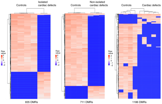
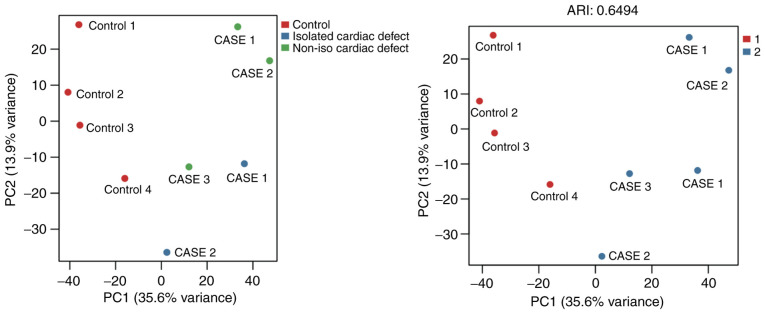
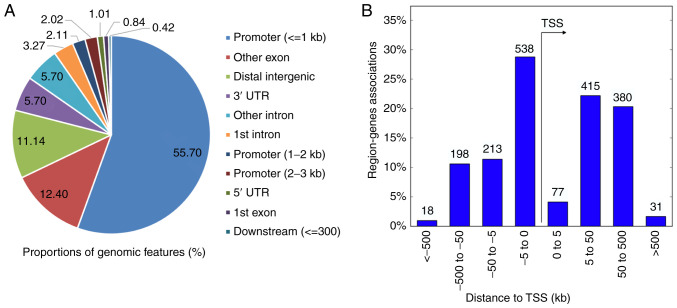
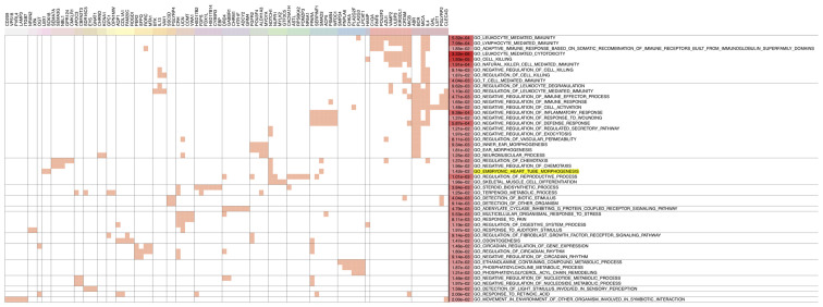
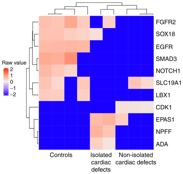
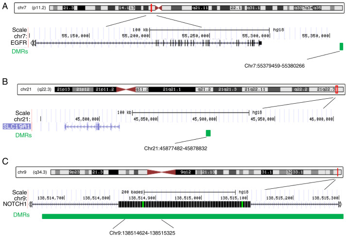
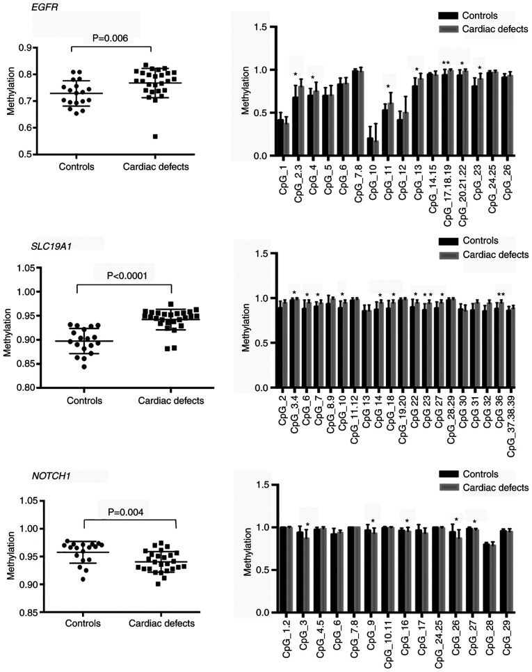
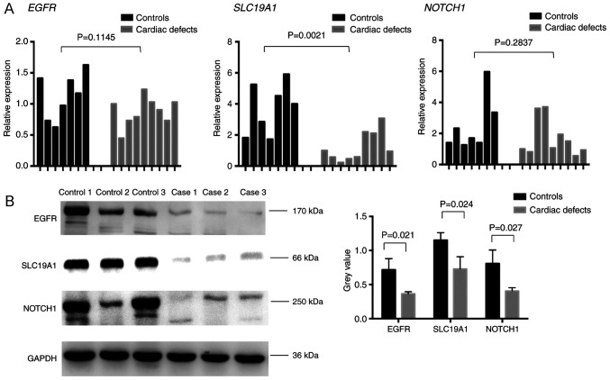
DNA methylation, as an epigenetic mechanism, has a vital role in heart development. An increasing number of studies have investigated aberrant DNA methylation in pediatric or adult heart samples from patients with congenital heart defects (CHD). Placenta tissue, umbilical cord blood, or newborn blood have also been used to detect DNA methylation biomarkers for CHD. However, few studies have compared the methylation levels in fetal heart tissue with cardiac defects with that in normal controls. The present study conducted an integrative whole-genome and CpG site-specific DNA methylation analysis of fetal heart samples from 17 isolated cardiac defect cases, 14 non-isolated cardiac defect cases, and 22 controls with normal hearts, using methylated DNA immunoprecipitation microarray and MassARRAY EpiTYPER assays. Expression of genes adjacent to differentially methylated regions (DMRs) was measured by RT-qPCR and western blot analysis. The results revealed that fetuses with cardiac defects presented global hypomethylation. Genomic analysis of DMRs revealed that a proportion of DMRs were located in exons (12.4%), distal intergenic regions (11.14%), and introns (8.97%). Only 55.7% of DMRs were observed at promoter regions. Functional enrichment analysis for genes adjacent to these DMRs revealed that hypomethylated genes were involved in embryonic heart tube morphogenesis and immune-related regulation functions. Intergenic hypermethylation of EGFR and solute carrier family 19 member 1 (SLC19A1), and intragenic hypomethylation of NOTCH1 were validated in fetal heart tissues with cardiac defects. Only SLC19A1 expression was significantly decreased at the mRNA level, while EGFR, NOTCH1, and SLC19A1 expression were all significantly decreased at the protein level. In conclusion, the present study demonstrated that fetal cardiac defects may be associated with alterations in regional and single CpG site methylation outside of promoter regions, resulting in differentiated expression of corresponding genes associated with heart development. These results present new insights into the epigenetic mechanisms underlying abnormal heart development.

Keywords: CpG site methylation; cardiac defects; fetal; genes expression alterations.


Conflict of interest statement

The authors declare that they have no competing interests.

Figures

Figure 1
Figure 1
Heatmaps of DMRs showing relative hypomethylation levels in fetal heart with isolated cardiac defects, non-isolated cardiac defects, and cardiac defects irrespective of whether they are isolated. DMRs, differentially methylated regions.
Figure 2
Figure 2
Principal component analysis for the most variable methylated levels of 1,737 CpG sites. The red dots represent the normal controls. The blue and green dots represent samples with cardiac defects. PC, principal component; ARI, Adjusted Rand index.
Figure 3
Figure 3
Genomic distribution of 1,186 DMRs across the genome. (A) The pie chart shows the proportions of genomic features (%). (B) The histogram shows the locations of DMRs in relation to the TSS. DMRs, differentially methylated regions; TSS, transcription start site; UTR, untranslated region.
Figure 4
Figure 4
Functional enrichment analysis of 1,052 hypomethylated differentially methylated regions related to annotated genes in fetal heart with cardiac defects. Each row represents one GO term. Each column represents one gene. The GO term ‘embryonic heart tube morphogenesis’ is highlighted in yellow. GO, gene ontology.
Figure 5
Figure 5
Heatmap of 11 differentially methylated regions corresponding to annotated genes which are associated with heart development based on enrichment analysis. FGFR2, fibroblast growth factor receptor 2; SOX18, SRY-box transcription factor 18; EGFR, epidermal growth factor receptor; SLC19A1, solute carrier family 19 member 1; LBX1, ladybird homeobox 1; CDK1, cyclin-dependent kinase 1; EPAS1, endothelial PAS domain protein 1; NPFF, neuropeptide FF-amide peptide precursor; ADA, adenosine deaminase.
Figure 6
Figure 6
Detailed schematic of CpG sites located in DMRs as summarized by DNA methylation levels in the downstream regions of (A) EGFR and (B) SLC19A1, and (C) in the exonic region of NOTCH1. DMRs, differentially methylated regions; EGFR, epidermal growth factor receptor; SLC19A1, solute carrier family 19 member 1.
Figure 7
Figure 7
Percentage methylation comparisons between controls (n=18) and cases with cardiac defects (n=26) at differentially methylated regions corresponding to EGFR, SLC19A1, and NOTCH1. Columns represent mean percentage methylation values, and the error bars represent standard deviation. *P<0.05 and **P<0.001. EGFR, epidermal growth factor receptor; SLC19A1, solute carrier family 19 member 1.
Figure 8
Figure 8
Validation of EGFR, SLC19A1, and NOTCH1 expression. (A) mRNA expression levels were examined by reverse transcription-quantitative PCR in single fetal hearts from seven normal controls (black bars) and nine cases with cardiac defects (gray bars). (B) Representative images and quantification of protein expression levels as determined by western blot analysis. EGFR, epidermal growth factor receptor; SLC19A1, solute carrier family 19 member 1.

References

    1. Hoffman JI, Kaplan S. The incidence of congenital heart disease. J Am Coll Cardiol. 2002;39:1890–1900. doi: 10.1016/s0735-1097(02)01886-7. - DOI - PubMed
    1. Yuan S, Zaidi S, Brueckner M. Congenital heart disease: Emerging themes linking genetics and development. Curr Opin Genet Dev. 2013;23:352–359. doi: 10.1016/j.gde.2013.05.004. - DOI - PMC - PubMed
    1. Vecoli C, Pulignani S, Foffa I, Andreassi MG. Congenital heart disease: The crossroads of genetics, epigenetics and environment. Curr Genomics. 2014;15:390–399. doi: 10.2174/1389202915666140716175634. - DOI - PMC - PubMed
    1. Jarrell DK, Lennon ML, Jacot JG. Epigenetics and mechanobiology in heart development and congenital heart disease. Diseases. 2019;7(52) doi: 10.3390/diseases7030052. - DOI - PMC - PubMed
    1. Greenberg MVC, Bourc'his D. The diverse roles of DNA methylation in mammalian development and disease. Nat Rev Mol Cell Biol. 2019;20:590–607. doi: 10.1038/s41580-019-0159-6. - DOI - PubMed