. 2021 Aug;14(8):e008170.
doi: 10.1161/CIRCHEARTFAILURE.120.008170. Epub 2021 Aug 10.

NAD+ Redox Imbalance in the Heart Exacerbates Diabetic Cardiomyopathy

Affiliations

NAD+ Redox Imbalance in the Heart Exacerbates Diabetic Cardiomyopathy

Ying Ann Chiao et al. Circ Heart Fail. 2021 Aug.

Abstract

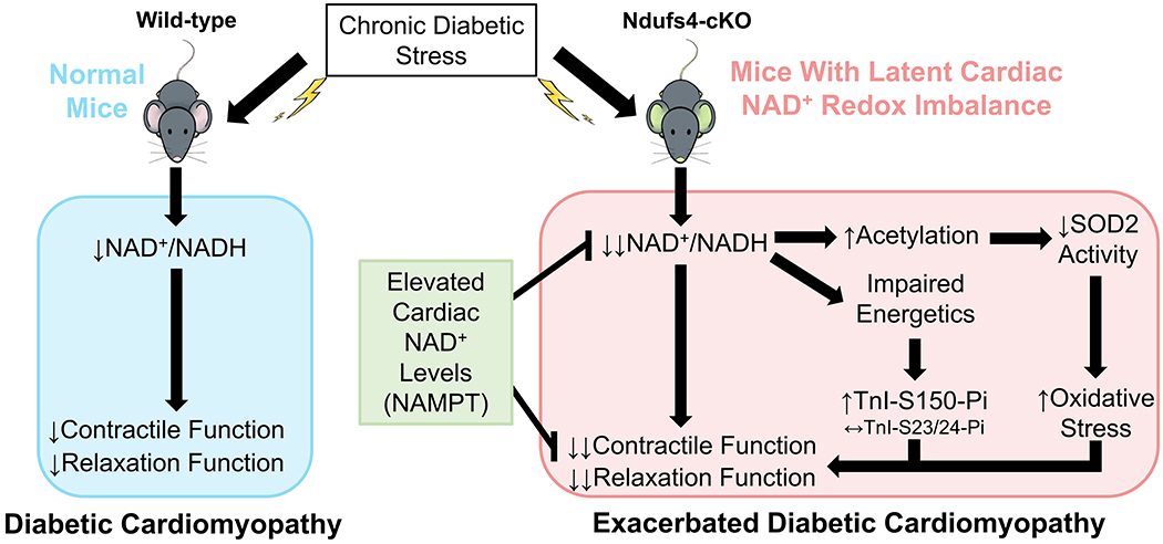
Background: Diabetes is a risk factor for heart failure and promotes cardiac dysfunction. Diabetic tissues are associated with nicotinamide adenine dinucleotide (NAD+) redox imbalance; however, the hypothesis that NAD+ redox imbalance causes diabetic cardiomyopathy has not been tested. This investigation used mouse models with altered NAD+ redox balance to test this hypothesis.

Methods: Diabetic stress was induced in mice by streptozotocin. Cardiac function was measured by echocardiography. Heart and plasma samples were collected for biochemical, histological, and molecular analyses. Two mouse models with altered NAD+ redox states (1, Ndufs4 [NADH:ubiquinone oxidoreductase subunit S4] knockout, cKO, and 2, NAMPT [nicotinamide phosphoribosyltranferase] transgenic mice, NMAPT) were used.

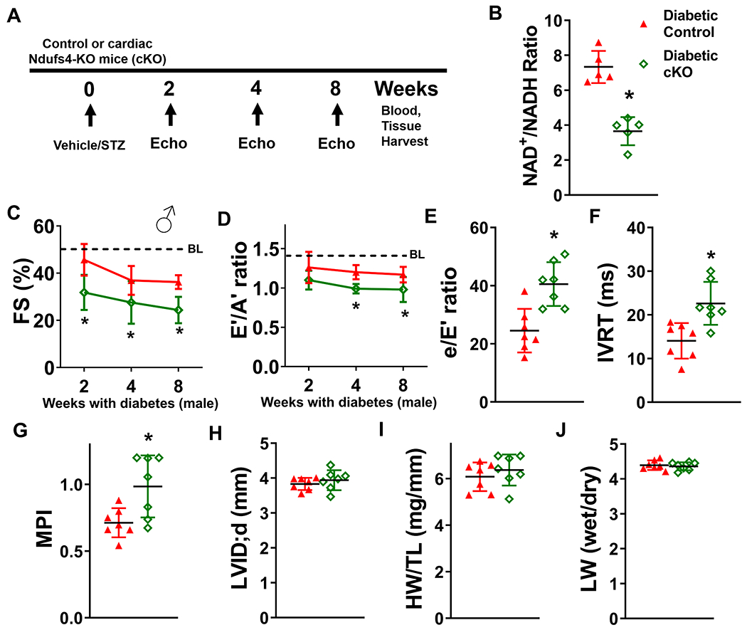
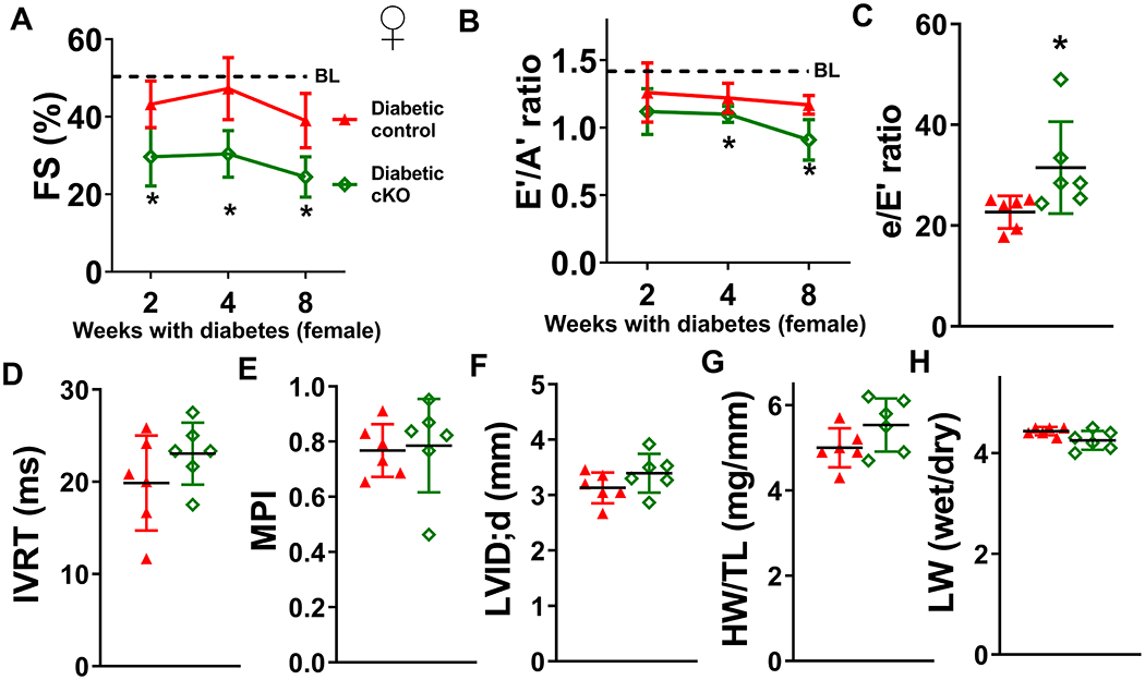
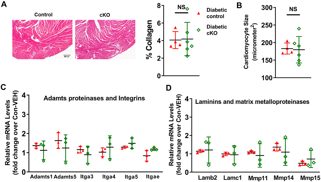
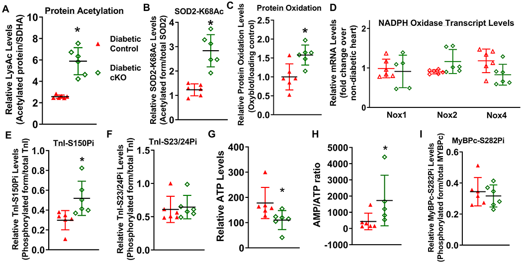
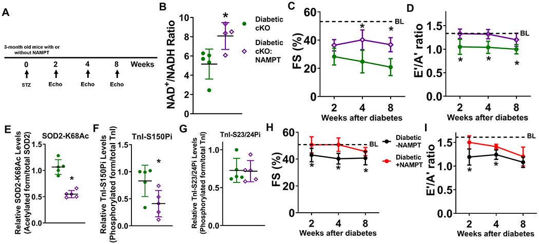
Results: Diabetic stress caused cardiac dysfunction and lowered NAD+/NADH ratio (oxidized/reduced ratio of nicotinamide adenine dinucleotide) in wild-type mice. Mice with lowered cardiac NAD+/NADH ratio without baseline dysfunction, cKO mice, were challenged with chronic diabetic stress. NAD+ redox imbalance in cKO hearts exacerbated systolic (fractional shortening: 27.6% versus 36.9% at 4 weeks, male cohort P<0.05), and diastolic dysfunction (early-to-late ratio of peak diastolic velocity: 0.99 versus 1.20, P<0.05) of diabetic mice in both sexes. Collagen levels and transcripts of fibrosis and extracellular matrix-dependent pathways did not show changes in diabetic cKO hearts, suggesting that the exacerbated cardiac dysfunction was due to cardiomyocyte dysfunction. NAD+ redox imbalance promoted superoxide dismutase 2 acetylation, protein oxidation, troponin I S150 phosphorylation, and impaired energetics in diabetic cKO hearts. Importantly, elevation of cardiac NAD+ levels by NAMPT normalized NAD+ redox balance, alleviated cardiac dysfunction (fractional shortening: 40.2% versus 24.8% in cKO:NAMPT versus cKO, P<0.05; early-to-late ratio of peak diastolic velocity: 1.32 versus 1.04, P<0.05), and reversed pathogenic mechanisms in diabetic mice.

Conclusions: Our results show that NAD+ redox imbalance to regulate acetylation and phosphorylation is a critical mediator of the progression of diabetic cardiomyopathy and suggest the therapeutic potential for diabetic cardiomyopathy by harnessing NAD+ metabolism.

Keywords: NAD+ redox imbalance; cardiomyopathy; diabetes; heart failure; risk factor.


Conflict of interest statement

Disclosures

No financial or non-financial competing interest is disclosed associated with the investigation in this manuscript by all authors.

Figures

Figure 1.
Figure 1.. 16-week diabetic stress leads to systolic and diastolic dysfunction and lowered NAD+/NADH ratio.
C57/BL6 wild type (WT) mice were treated with STZ to induce diabetes. Cardiac function was assessed with echocardiography 16 weeks after induction of diabetes. (A) Plasma glucose levels were measured 16 weeks after vehicle or STZ treatment. (B) Fractional shortening (FS), (C) E’/A’, (D) e/E’ ratio, and (E) isovolumic relaxation time (IVRT) were measured to evaluate systolic and diastolic functions. (F) Representative pulsed-wave (upper panels) and tissue (middle panels) doppler images and corresponding electrocardiogram (lower panels) images. (G) Cardiac NAD+/NADH ratio were measured. N=6. *: P<0.05 to vehicle. (H) FS, (I) E’/A’ ratio, (J) e/E’ ratio, (K) IVRT and (L) cardiac NAD+/NADH ratio were measured in mice 2-week after diabetic stress. N=6. *: P<0.05 to vehicle.
Figure 2.
Figure 2.. Latent NAD(H) redox imbalance in the heart exacerbates cardiac dysfunctions of diabetic male mice.
(A) Experimental plan. Control and cKO male mice were treated with STZ to induce diabetes. Cardiac function was measured at 2, 4, and 8 weeks after induction of diabetes. Blood and tissue samples were collected at the endpoint of the study. (B) Cardiac NAD+/NADH ratio was measured. N=5. Longitudinal changes in (C) fractional shortening (FS) and (D) E’/A’ ratio of diabetic control or cKO male mice were assessed. Dotted lines indicate average baseline (BL) values of non-diabetic control mice. Numerical values are available in Supplementary Table I. (E) e/E’ ratio, (F) isovolumic relaxation time (IVRT), (G) myocardial performance index (MPI) and (H) left ventricular internal dimension at diastole (LVID;d) were measured at 8 weeks after diabetes induction. (I) Cardiac hypertrophy (heart weight/tibia length; HW/TL) and (J) lung edema of diabetic control or cKO mice (lung wet weight/lung dry weight ratio; LW wet/dry) were determined at 8-week endpoint. N=7. *: P<0.05 to diabetic control mice.
Figure 3.
Figure 3.. NAD(H) redox imbalance in the heart also exacerbates cardiac dysfunctions of diabetic female mice.
Control and cKO female mice were treated with STZ to induce diabetes. Cardiac function was assessed at 2, 4, and 8 weeks after induction of diabetes. (A) Longitudinal changes in fractional shortening (FS) and (B) E’/A’ ratio of diabetic control or cKO female mice were measured. Dotted lines indicate average baseline (BL) values of non-diabetic control mice. Numerical values are available in Supplementary Table I. (C) e/E’ ratio, (D) IVRT, (E) MPI, (F) LVID;d, (G) HW/TL, and (H) LW wet/dry ratio of diabetic control or cKO female mice were determined at 8-week endpoint. N=6. *: P<0.05 to diabetic control mice.
Figure 4.
Figure 4.. NAD+ redox imbalance exacerbates diabetic cardiomyopathy, independent on tissue fibrosis.
(A) Collagen levels of diabetic mouse hearts were quantified by trichrome staining. (B) Cardiomyocyte sizes of these hearts were quantified. N=4-6. Transcript expression levels of fibrosis-related genes were quantified by qPCR analyses. (C) Adamts proteinases and integrins, and (D) laminins and MMPs in diabetic control and diabetic cKO male hearts were measured. N=3.
Figure 5.
Figure 5.. NAD+ redox imbalance regulated SOD2 acetylation, protein oxidation and myofilament protein phosphorylation.
(A) Global lysine acetylation levels of protein extracts from diabetic control or diabetic cKO hearts were assessed by Western blot. (B) Acetylation levels of SOD2 at lysine-68 were assessed by Western blot analysis. (C) Protein oxidation levels of diabetic mouse hearts were determined by Oxyblot analysis. (D) Relative mRNA levels of pro-oxidant genes (Nox1, Nox2 and Nox4) were measured by qPCR. Phosphorylation levels of (E) TnI at Serine 150 (TnI-S150Pi), (F) TnI-S23/24Pi, (G) ATP levels and (H) AMP/ATP ratio were measured. Phosphorylation levels of (I) MyBPc at Serine 282 (MyBPc-S282Pi) were measured. N=6. *: P<0.05 to diabetic control mice.
Figure 6.
Figure 6.. Elevation of cardiac NAD+ levels alleviates diabetic cardiomyopathy in cKO and control mice.
(A) Experimental plan. STZ was administered to male cKO and cKO:NAMPT mice to induce diabetes. Cardiac function of cKO and cKO:NAMPT mice was measured at 2, 4, and 8 weeks after induction of diabetes. (B) Cardiac NAD+/NADH ratio were measured. N=4-5. (C) FS and (D) E’/A’ ratio were assessed longitudinally. N=6. Dotted lines indicate average baseline (BL) values of non-diabetic control mice. Numerical values are available in Supplementary Table I. Levels of (E) SOD2-K68Ac, (F) TnI-S150Pi, and (G) TnI-S23/24Pi of indicated hearts were measured by Western blots. N=5. (H) FS and (I) E’/A’ ratio were measured longitudinally in control mice with or without NAMPT expression in the hearts after diabetes induction. N=5. *: P<0.05 to diabetic cKO mice or diabetic-NAMPT mice.
Figure 7.
Figure 7.. Graphical summary.
This study revealed the causal roles of NAD+ redox imbalance and cardiac dysfunction in hearts under chronic diabetic stress. In a mouse model with latent NAD+ redox imbalance (Ndufs4-cKO), we observed exacerbation of cardiac dysfunction in responses to chronic diabetic stress. The exacerbated contractile and relaxation dysfunction was mediated by protein hyperacetylation, in particular SOD2 acetylation which led to increased oxidative stress, and increased phosphorylation of TnI-S150 triggered by impaired energetics. Importantly, cardiac-specific elevation of NAD+ levels ameliorated diabetic cardiomyopathy and reversed pathogenic mechanisms, supporting the roles of NAD+ redox imbalance in the progression of diabetic cardiomyopathy.

References

    1. Boudina S and Abel ED. Diabetic cardiomyopathy, causes and effects. Rev Endocr Metab Disord. 2010;11:31–9. - PMC - PubMed
    1. Ritchie RH and Abel ED. Basic Mechanisms of Diabetic Heart Disease. Circ Res. 2020;126:1501–1525. - PMC - PubMed
    1. Imai S and Guarente L. NAD+ and sirtuins in aging and disease. Trends Cell Biol. 2014;24:464–71. - PMC - PubMed
    1. Guarente L Calorie restriction and sirtuins revisited. Genes Dev. 2013;27:2072–85. - PMC - PubMed
    1. Choudhary C, Weinert BT, Nishida Y, Verdin E and Mann M. The growing landscape of lysine acetylation links metabolism and cell signalling. Nature reviews Molecular cell biology. 2014;15:536–50. - PubMed

Publication types

MeSH terms