. 2021 Dec;301(3):E419-E425.
doi: 10.1148/radiol.2021211162. Epub 2021 Aug 10.

Cardiac MRI in Patients with Prolonged Cardiorespiratory Symptoms after Mild to Moderate COVID-19

Affiliations

Cardiac MRI in Patients with Prolonged Cardiorespiratory Symptoms after Mild to Moderate COVID-19

Dmitrij Kravchenko et al. Radiology. 2021 Dec.

Abstract

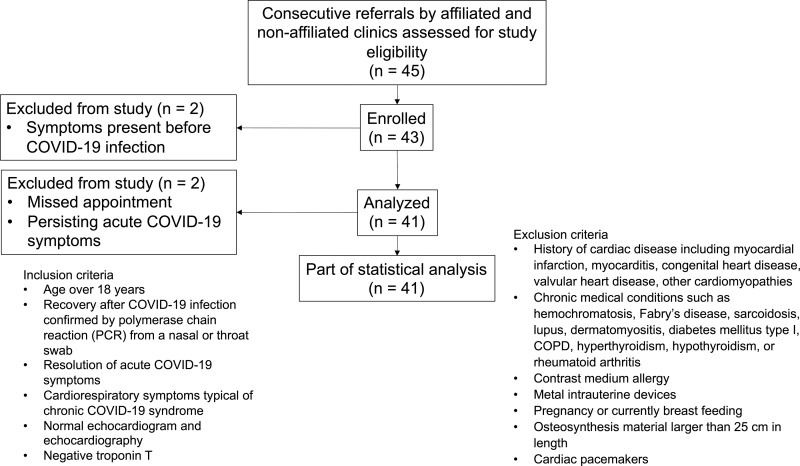
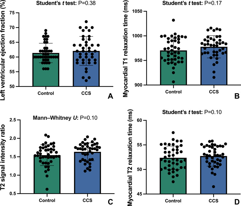
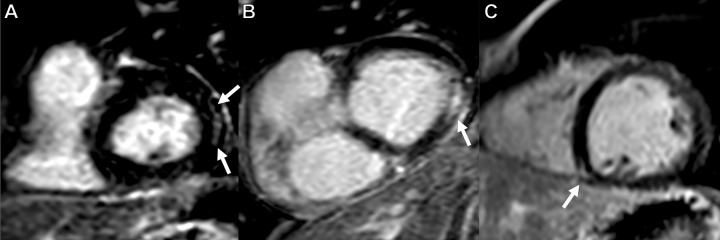
Background Myocardial injury and inflammation at cardiac MRI in patients with COVID-19 have been described in recent publications. Concurrently, a chronic COVID-19 syndrome (CCS) after SARS-CoV-2 infection has been observed and manifests with symptoms such as fatigue and exertional dyspnea. Purpose To explore the relationship between CCS and myocardial injury and inflammation as an underlying cause of the persistent complaints in previously healthy individuals. Materials and Methods In this prospective study from January 2021 to April 2021, study participants without known cardiac or pulmonary diseases prior to SARS-CoV-2 infection who had persistent CCS symptoms such as fatigue or exertional dyspnea after convalescence and healthy control participants underwent cardiac MRI. The cardiac MRI protocol included evaluating the T1 and T2 relaxation times, extracellular volume, T2 signal intensity ratio, and late gadolinium enhancement (LGE). Student t tests, Mann-Whitney U tests, and χ2 tests were used for statistical analysis. Results Forty-one participants with CCS (mean age, 39 years ± 13 [standard deviation]; 18 men) and 42 control participants (mean age, 39 years ± 16; 26 men) were evaluated. The median time between the initial incidence of mild to moderate COVID-19 not requiring hospitalization and undergoing cardiac MRI was 103 days (interquartile range, 88-158 days). Troponin T levels were normal. Parameters indicating myocardial inflammation and edema were comparable between participants with CCS and control participants (T1 relaxation times: 978 msec ± 23 vs 971 msec ± 25 [P = .17]; T2 relaxation times: 53 msec ± 2 vs 52 msec ± 2 [P = .47]; T2 signal intensity ratios: 1.6 ± 0.2 vs 1.6 ± 0.3 [P = .10]). Visible myocardial edema was present in none of the participants. Three of 41 (7%) participants with CCS demonstrated nonischemic LGE, whereas no participants in the control group demonstrated nonischemic LGE (0 of 42 [0%]; P = .07). None of the participants fulfilled the 2018 Lake Louise criteria for the diagnosis of myocarditis. Conclusion Individuals with chronic COVID-19 syndrome who did not undergo hospitalization for COVID-19 did not demonstrate signs of active myocardial injury or inflammation at cardiac MRI. © RSNA, 2021 Online supplemental material is available for this article. See also the editorial by Lima and Bluemke in this issue.


Conflict of interest statement

Disclosures of Conflicts of Interest: D. Kravchenko Stocks in Pfizer, Johnson & Johnson, and Abvvie. A.I. No relevant relationships. S.Z. No relevant relationships. N.M. No relevant relationships. M.R. No relevant relationships. A.F. No relevant relationships. C.C.P. Payment or honoraria for lectures, presentations, speakers bureaus, manuscript writing or educational events from Guerbet, Bayer, Vital, Philips Healthcare, and Julius Zorn. A.H. No relevant relationships. M.V. No relevant relationships. J.N. No relevant relationships. D. Kuetting No relevant relationships. G.D.D. No relevant relationships. U.I.A. Payment or honoraria for lectures, presentations, speakers bureaus, manuscript writing or educational events and support for attending meetings and/or travel from Siemens Healthineers. J.A.L. Payment or honoraria for lectures, presentations, speakers bureaus, manuscript writing or educational events from Philips Healthcare.

Figures

Flowchart depicts the recruitment process, inclusion and exclusion criteria, and included participants with chronic COVID-19 syndrome. COPD = chronic obstructive pulmonary disease, PCR = polymerase chain reaction.
Figure 1:
Flowchart depicts the recruitment process, inclusion and exclusion criteria, and included participants with chronic COVID-19 syndrome. COPD = chronic obstructive pulmonary disease, PCR = polymerase chain reaction.
Column graphs with individual plotted values show the distribution of MRI parameters in the control group and in the group with chronic COVID-19 syndrome (CCS). The means of the data are represented by bars. Whiskers represent standard deviations. The distribution is shown for the (A) left ventricular ejection fraction, (B) myocardial T1 relaxation time, (C) T2 signal intensity ratio, and (D) myocardial T2 relaxation time.
Figure 2:
Column graphs with individual plotted values show the distribution of MRI parameters in the control group and in the group with chronic COVID-19 syndrome (CCS). The means of the data are represented by bars. Whiskers represent standard deviations. The distribution is shown for the (A) left ventricular ejection fraction, (B) myocardial T1 relaxation time, (C) T2 signal intensity ratio, and (D) myocardial T2 relaxation time.
Late gadolinium enhancement (LGE) short-axis views demonstrate LGE in three of 41 patients (7%). Subepicardial LGE along the basal inferolateral wall (arrows) in (A) a 63-year-old man and (B) a 54-year-old man. (C) LGE at the right ventricular attachment (arrow) in a 19-year-old man.
Figure 3:
Late gadolinium enhancement (LGE) short-axis views demonstrate LGE in three of 41 patients (7%). Subepicardial LGE along the basal inferolateral wall (arrows) in (A) a 63-year-old man and (B) a 54-year-old man. (C) LGE at the right ventricular attachment (arrow) in a 19-year-old man.

Comment in

References

    1. Gavriatopoulou M , Korompoki E , Fotiou D , et al. . Organ-specific manifestations of COVID-19 infection . Clin Exp Med 2020. ; 20 ( 4 ): 493 – 506 . - PMC - PubMed
    1. Chang WT , Toh HS , Liao CT , Yu WL . Cardiac involvement of COVID-19: a comprehensive review . Am J Med Sci 2021. ; 361 ( 1 ): 14 – 22 . - PMC - PubMed
    1. Gupta A , Madhavan MV , Sehgal K , et al. . Extrapulmonary manifestations of COVID-19 . Nat Med 2020. ; 26 ( 7 ): 1017 – 1032 . - PMC - PubMed
    1. Townsend L , Dyer AH , Jones K , et al. . Persistent fatigue following SARS-CoV-2 infection is common and independent of severity of initial infection . PLoS One 2020. ; 15 ( 11 ): e0240784 . - PMC - PubMed
    1. Wostyn P . COVID-19 and chronic fatigue syndrome: Is the worst yet to come? Med Hypotheses 2021. ; 146 110469 . - PMC - PubMed

Publication types