

Prenatal genetic diagnosis of omphalocele by karyotyping, chromosomal microarray analysis and exome sequencing
- PMID: 34374610
- PMCID: PMC8366676
- DOI: 10.1080/07853890.2021.1962966
Prenatal genetic diagnosis of omphalocele by karyotyping, chromosomal microarray analysis and exome sequencing
Abstract
Objectives: The aim of this study is to share our experience in the prenatal diagnosis of omphalocele by karyotyping, chromosomal microarray analysis (CMA) and whole exome sequencing (WES).
Methods: In this retrospective study, 81 cases of omphalocele were identified from 2015 to 2020. Associated anomalies and prenatal diagnosis based on karyotyping, CMA and WES were analysed.
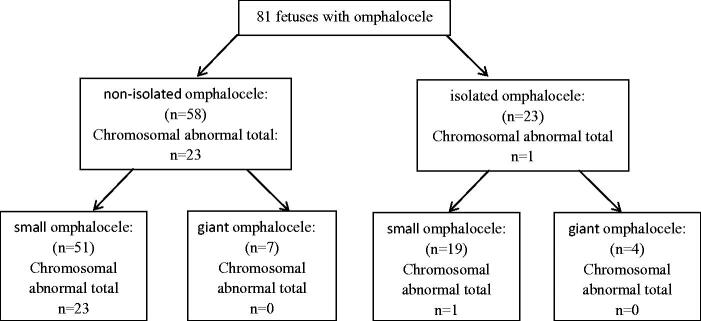
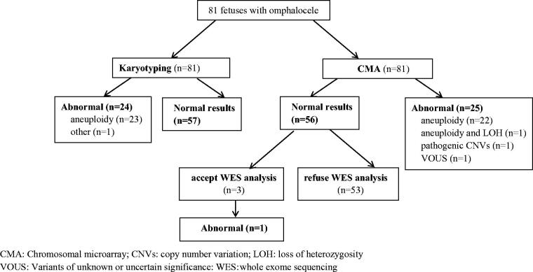
Results: Fifty-eight (71.6%) of the 81 foetuses had other ultrasound anomalies. Giant omphalocele was present in 11 cases (13.6%) and small omphalocele was present in 70 cases (86.4%). Chromosomal abnormalities were found in 24 foetuses (29.6%, 24/81), the most common of which were trisomy 18 (58.8%, 11/24) and trisomy 13 (29.2%, 7/24). Compared to isolated omphalocele, non-isolated omphalocele was accompanied by an increased prevalence of chromosomal abnormalities (4.3% (1/23) vs. 39.7% (23/58), χ2 = 8.226, p = .004). All chromosomal abnormalities were found in small omphalocele. Aside from aneuploidy, CMA showed one pathogenic copy number variants (CNVs) for a detection rate of 1.2%, one variants of unknown significance (VOUS) and one instance of loss of heterozygosity (LOH). WES was performed on 3 non-isolated cases, and one was found to have pathogenic variants.
Conclusions: The most common genetic cause of omphalocele is aneuploidy and the prevalence of chromosomal abnormalities is increased with non-isolated and small omphalocele. CMA and WES can be useful for providing further genetic information to assist in prenatal counselling and pregnancy management.
Keywords: Omphalocele; chromosomal microarray analysis; karyotyping; prenatal diagnosis; whole-exome sequencing.
Conflict of interest statement
The authors have no conflicts to declare.
Figures
References
-
- Calvert N, Damiani S, Sunario J, et al. The outcomes of pregnancies following a prenatal diagnosis of fetal exomphalos in Western Australia. Aust N Z J Obstet Gynaecol. 2009;49(4):371–375. - PubMed
-
- Verla MA, Style CC, Olutoye OO.. Prenatal diagnosis and management of omphalocele. Semin Pediatr Surg. 2019;28(2):84–88. - PubMed
-
- Adams AD, Stover S, Rac MW.. Omphalocele-What should we tell the prospective parents? Prenat Diagn. 2021;41(4):486–496. - PubMed
-
- Tong H, Lu J, Liu L, et al. Prenatal diagnosis of suspected recurrent Beckwith-Wiedeman syndrome:a case report and literature review. Chin J Perinat Med. 2021;24(4):283–287.
Publication types
MeSH terms
LinkOut - more resources
Full Text Sources
Medical