Comparative Study
. 2021 Nov 15;204(10):1164-1179.
doi: 10.1164/rccm.202104-0847OC.

Diagnostic Accuracy of Endobronchial Optical Coherence Tomography for the Microscopic Diagnosis of Usual Interstitial Pneumonia

Affiliations
Comparative Study

Diagnostic Accuracy of Endobronchial Optical Coherence Tomography for the Microscopic Diagnosis of Usual Interstitial Pneumonia

Sreyankar Nandy et al. Am J Respir Crit Care Med. .

Abstract

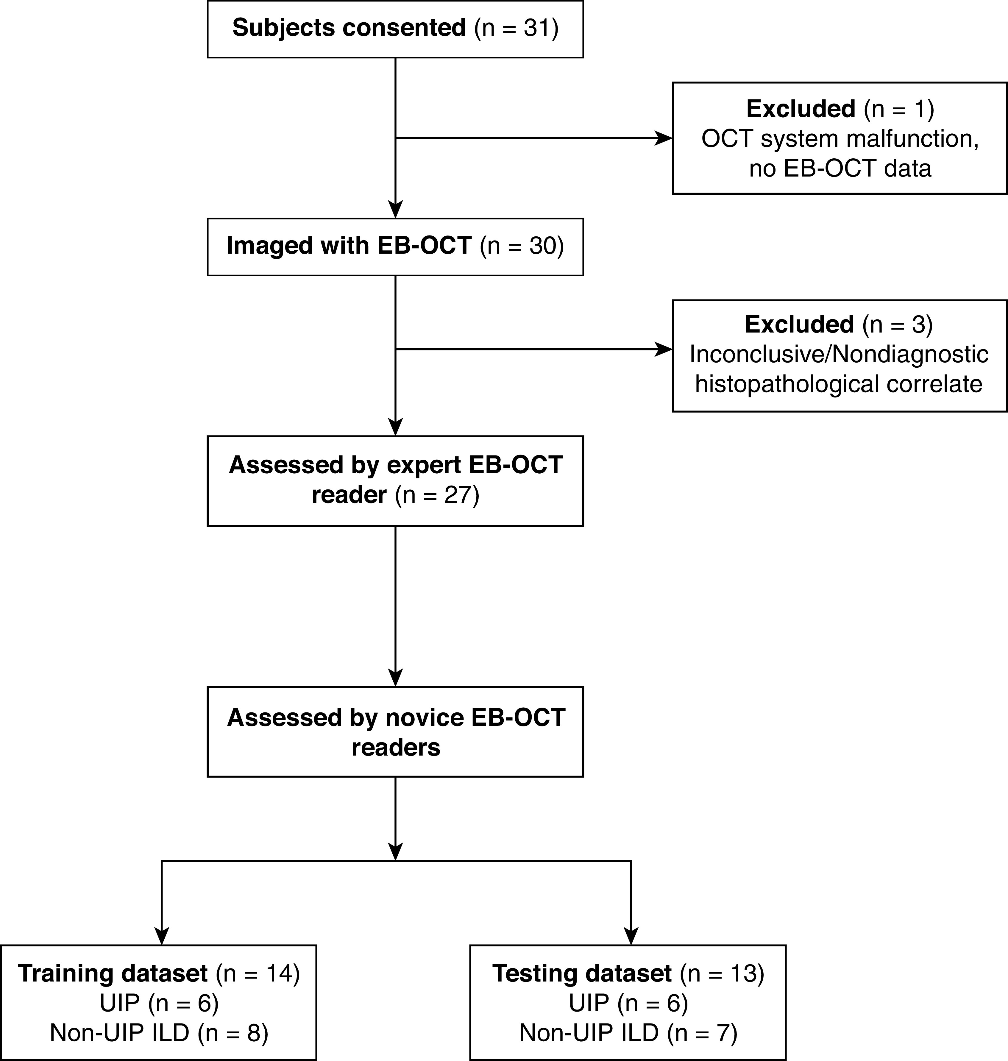
Rationale: Early, accurate diagnosis of interstitial lung disease (ILD) informs prognosis and therapy, especially in idiopathic pulmonary fibrosis (IPF). Current diagnostic methods are imperfect. High-resolution computed tomography has limited resolution, and surgical lung biopsy (SLB) carries risks of morbidity and mortality. Endobronchial optical coherence tomography (EB-OCT) is a low-risk, bronchoscope-compatible modality that images large lung volumes in vivo with microscopic resolution, including subpleural lung, and has the potential to improve the diagnostic accuracy of bronchoscopy for ILD diagnosis. Objectives: We performed a prospective diagnostic accuracy study of EB-OCT in patients with ILD with a low-confidence diagnosis undergoing SLB. The primary endpoints were EB-OCT sensitivity/specificity for diagnosis of the histopathologic pattern of usual interstitial pneumonia (UIP) and clinical IPF. The secondary endpoint was agreement between EB-OCT and SLB for diagnosis of the ILD fibrosis pattern. Methods: EB-OCT was performed immediately before SLB. The resulting EB-OCT images and histopathology were interpreted by blinded, independent pathologists. Clinical diagnosis was obtained from the treating pulmonologists after SLB, blinded to EB-OCT. Measurements and Main Results: We enrolled 31 patients, and 4 were excluded because of inconclusive histopathology or lack of EB-OCT data. Twenty-seven patients were included in the analysis (16 men, average age: 65.0 yr): 12 were diagnosed with UIP and 15 with non-UIP ILD. Average FVC and DlCO were 75.3% (SD, 18.5) and 53.5% (SD, 16.4), respectively. Sensitivity and specificity of EB-OCT was 100% (95% confidence interval, 75.8-100.0%) and 100% (79.6-100%), respectively, for both histopathologic UIP and clinical diagnosis of IPF. There was high agreement between EB-OCT and histopathology for diagnosis of ILD fibrosis pattern (weighted κ: 0.87 [0.72-1.0]). Conclusions: EB-OCT is a safe, accurate method for microscopic ILD diagnosis, as a complement to high-resolution computed tomography and an alternative to SLB.

Keywords: idiopathic pulmonary fibrosis; interstitial lung disease; in vivo microscopy; in vivo optical imaging; usual interstitial pneumonia.


Figures

Figure 1.
Figure 1.
Flow diagram of study enrollment. EB-OCT = endobronchial optical coherence tomography; ILD = interstitial lung disease; UIP = usual interstitial pneumonia.
Figure 2.
Figure 2.
EB-OCT of alveolar lung parenchyma. (A) In vivo volumetric EB-OCT and (B and C) corresponding cross-sectional EB-OCT images from the upper lobe show alveolated lung parenchyma with minimal fibrosis. Alveoli appear as round, evenly spaced and sized, signal-void (black) structures with thin, lattice-like alveolar walls attached to the thin distal bronchiolar wall. (D) Representative histology from the surgical lung biopsy confirms regions of preserved lung parenchyma with minimal interstitial fibrosis. The EB-OCT cross-sections in B and C are taken from the locations on the volumetric image (A) indicated by the blue and yellow squares, respectively. a = alveoli; aw = alveolar walls; EB-OCT = endobronchial optical coherence tomography.
Figure 3.
Figure 3.
EB-OCT of UIP in a patient ultimately diagnosed with histopathologic UIP and clinical IPF. (A) In vivo volumetric EB-OCT and (B and C) corresponding cross-sectional EB-OCT images from the lower lobe of the same patient in Figure 2 show features of UIP. Dense, signal-intense (light gray to white) subpleural fibrosis has obliterated alveolar structures. Embedded within the fibrosis are clusters of enlarged, irregularly shaped, stacked, signal-poor (black to dark gray) cystic structures, consistent with microscopic honeycombing. (D) Subsequent surgical lung biopsy confirms dense, destructive subpleural fibrosis with infrequent, individual cystic honeycomb-like structures. The consensus histopathologic diagnosis was UIP, but there was wide discrepancy among the reviewing pathologists, owing primarily to the absence of honeycombing. The diagnosis of UIP was ultimately confirmed 3 years later on surgical explant lung histology at the time of lung transplantation, and the patient was clinically diagnosed with IPF. The EB-OCT cross-sections in B and C are taken from the locations on the volumetric image (A) indicated by the blue and yellow squares, respectively. EB-OCT = endobronchial optical coherence tomography; F = fibrosis; HC = microscopic honeycombing; IPF = idiopathic pulmonary fibrosis; UIP = usual interstitial pneumonia.
Figure 4.
Figure 4.
EB-OCT of UIP in a patient with combined pulmonary fibrosis and emphysema. (A) In vivo volumetric and (B and C) cross-sectional EB-OCT images from the basilar segment of the lower lobe show features of UIP, including microscopic honeycombing embedded in a background of dense, destructive subpleural fibrosis. A region of traction bronchiectasis can be seen as an abnormally dilated, tortuous airway branch point with fibrosis and some adjacent preserved alveolar spaces. (D) Corresponding surgical lung biopsy shows dense destructive fibrosis with microscopic honeycombing. The clinical follow-up diagnosis was idiopathic pulmonary fibrosis with emphysema, also known as combined pulmonary fibrosis and emphysema. The EB-OCT cross-sections in B and C are taken from the locations on the volumetric image (A) indicated by the blue and yellow squares, respectively. a = preserved alveolar spaces; EB-OCT = endobronchial optical coherence tomography; F = fibrosis; HC = microscopic honeycombing; TB = traction bronchiectasis; UIP = usual interstitial pneumonia.
Figure 5.
Figure 5.
EB-OCT of emphysema in a patient with combined pulmonary fibrosis and emphysema. (A) In vivo volumetric and (B and C) cross-sectional EB-OCT images from the superior segment of the lower lobe in the same patient as Figure 4 show emphysema, demonstrated as irregularly shaped, abnormally enlarged airspaces with focal destruction of alveolar walls. Fibrosis was minimal. (D) Corresponding surgical lung biopsy confirms the presence of emphysema. The emphysematous changes are distinguishable from usual interstitial pneumonia seen in the patient’s lower lobe basilar segment (Figure 4). The clinical follow-up diagnosis was idiopathic pulmonary fibrosis with emphysema, also known as combined pulmonary fibrosis and emphysema. The EB-OCT cross-sections in B and C are taken from the locations on the volumetric image (A) indicated by the blue and yellow squares, respectively. CPFE = combined pulmonary fibrosis and emphysema; ea = enlarged airspaces; EB-OCT = endobronchial optical coherence tomography.
Figure 6.
Figure 6.
EB-OCT in a patient with NSIP. (A) In vivo volumetric and (B and C) cross-sectional EB-OCT images show preserved alveolar architecture with mild, homogeneous fibrotic thickening of the interstitial alveolar walls, consistent with NSIP. A small normal bronchiolar airway branch point can also be seen. (D) Subsequent surgical lung biopsy confirms homogeneous interstitial fibrosis and the diagnosis of NSIP. The EB-OCT cross-sections in B and C are taken from the locations on the volumetric image (A) indicated by the blue and yellow squares, respectively. b = bronchiolar airway branch point; EB-OCT = endobronchial optical coherence tomography; IF = interstitial fibrosis in alveolar walls; NSIP = nonspecific interstitial pneumonia.
Figure 7.
Figure 7.
EB-OCT in a patient with airway-centered fibrosis. (A) In vivo volumetric and (B and C) cross-sectional EB-OCT from the lower lobe of a patient shows dense, signal-intense fibrosis surrounding the proximal end of the airway (left side). Traction bronchiectasis within the fibrotic region appears as a cystically dilated, irregular airway branch point. Adjacent, smaller, irregular cystic structures are indicative of peribronchiolar metaplasia, often associated with traction bronchiectasis in airway-centered fibrosis. Distal to the airway-centered fibrosis, there is a transition to alveolated parenchyma with enlarged, rounded airspaces and intact alveolar walls, indicative of hyperinflation. (D) Surgical lung biopsy confirmed the diagnosis of airway-centered fibrosis with traction bronchiectasis, peribronchiolar metaplasia, and enlarged airspaces with intact alveolar walls. The EB-OCT cross-sections in B and C are taken from the locations on the volumetric image (A) indicated by the blue and yellow squares, respectively. ea = enlarged, rounded airspaces; EB-OCT = endobronchial optical coherence tomography; F = fibrosis; PBM = peribronchiolar metaplasia; TB = traction bronchiectasis.
Figure 8.
Figure 8.
Flow diagram of potential incorporation of EB-OCT in clinical ILD diagnostic workflow. EB-OCT = endobronchial optical coherence tomography; HRCT = high-resolution computed tomography; ILD = interstitial lung disease; IPF = idiopathic pulmonary fibrosis; SLB = surgical lung biopsy; TBLC = transbronchial lung cryobiopsy; UIP = usual interstitial pneumonia.

Comment in

References

    1. Daniil ZD, Gilchrist FC, Nicholson AG, Hansell DM, Harris J, Colby TV, et al. A histologic pattern of nonspecific interstitial pneumonia is associated with a better prognosis than usual interstitial pneumonia in patients with cryptogenic fibrosing alveolitis. Am J Respir Crit Care Med . 1999;160:899–905. - PubMed
    1. Raghu G, Remy-Jardin M, Myers JL, Richeldi L, Ryerson CJ, Lederer DJ, et al. American Thoracic Society, European Respiratory Society, Japanese Respiratory Society, and Latin American Thoracic Society. Diagnosis of idiopathic pulmonary fibrosis: an official ATS/ERS/JRS/ALAT clinical practice guideline. Am J Respir Crit Care Med . 2018;198:e44–e68. - PubMed
    1. Lynch DA, Sverzellati N, Travis WD, Brown KK, Colby TV, Galvin JR, et al. Diagnostic criteria for idiopathic pulmonary fibrosis: a Fleischner Society white paper. Lancet Respir Med . 2018;6:138–153. - PubMed
    1. King TE, Jr, Bradford WZ, Castro-Bernardini S, Fagan EA, Glaspole I, Glassberg MK, et al. ASCEND Study Group. A phase 3 trial of pirfenidone in patients with idiopathic pulmonary fibrosis. N Engl J Med . 2014;370:2083–2092. - PubMed
    1. Richeldi L, du Bois RM, Raghu G, Azuma A, Brown KK, Costabel U, et al. INPULSIS Trial Investigators. Efficacy and safety of nintedanib in idiopathic pulmonary fibrosis. N Engl J Med . 2014;370:2071–2082. - PubMed

Publication types