

LncRNA-HAGLR motivates triple negative breast cancer progression by regulation of WNT2 via sponging miR-335-3p
- PMID: 34375306
- PMCID: PMC8386551
- DOI: 10.18632/aging.203272
LncRNA-HAGLR motivates triple negative breast cancer progression by regulation of WNT2 via sponging miR-335-3p
Abstract
Background: Triple negative breast cancer (TNBC) is a group of highly heterogeneous mixed breast cancer at the level of gene expression profile. Therefore, it is of great clinical significance to explore the molecular mechanism of TNBC and find a targeted therapeutic approach from the molecular level.
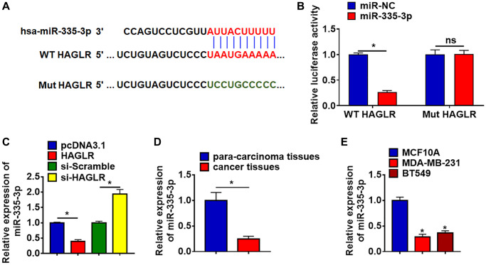
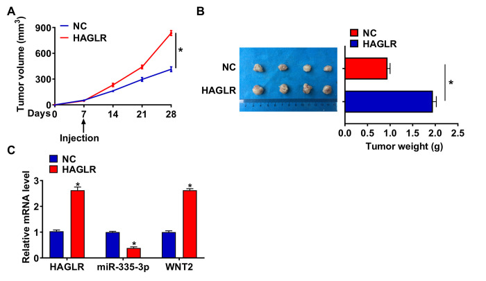
Methods: Long non-coding RNA (lncRNA) HAGLR expression level was measured by and qRT-PCR in TNBC tissues and cell lines. EdU, MTT, wound healing and Transwell assays were performed to explore the role of HAGLR on the malignancy of TNBC cells. Luciferase assay was used to clarify the binding between miR-335-3p with HAGLR and WNT2. The tumor formation experiment in nude mice was used to explore the function of HAGLR in vivo.
Results: HAGLR was increased in TNBC tissues and cell lines. Silencing of HAGLR inhibited viability, proliferation, migration, and invasion of BT549 cells. Furthermore, HAGLR acted as a sponge of miR-335-3p and inhibited its expression. And miR-335-3p directly targeted WNT2. Functionally, forced expression of miR-335-3p or knockdown of WNT2 removed the promoted effects of lncRNA HAGLR on TNBC development. In vivo tumorigenesis experiments indicated HAGLR accelerated tumor growth via miR-335-3p/WNT2 axis.
Conclusion: Our study revealed that HAGLR promoted the growth of TNBC, which was mediated by miR-335-3p/WNT2 axis.
Keywords: WNT2; lncRNA HAGLR; miR-335-3p; triple negative breast cancer; tumor progression.
Conflict of interest statement
Figures


Similar articles
-
A novel tumor suppressor ASMTL-AS1 regulates the miR-1228-3p/SOX17/β-catenin axis in triple-negative breast cancer.Diagn Pathol. 2021 May 18;16(1):45. doi: 10.1186/s13000-021-01105-3. Diagn Pathol. 2021. PMID: 34006305 Free PMC article.
-
Exploring the regulatory role of FBXL19-AS1 in triple-negative breast cancer through the miR-378a-3p/OTUB2 axis.Cell Biochem Funct. 2024 Jun;42(4):e4020. doi: 10.1002/cbf.4020. Cell Biochem Funct. 2024. PMID: 38702967
-
Long noncoding RNA Linc00339 promotes triple-negative breast cancer progression through miR-377-3p/HOXC6 signaling pathway.J Cell Physiol. 2019 Aug;234(8):13303-13317. doi: 10.1002/jcp.28007. Epub 2019 Jan 7. J Cell Physiol. 2019. PMID: 30618083
-
Long noncoding RNAs in triple-negative breast cancer: A new frontier in the regulation of tumorigenesis.J Cell Physiol. 2021 Dec;236(12):7938-7965. doi: 10.1002/jcp.30463. Epub 2021 Jun 8. J Cell Physiol. 2021. PMID: 34105151 Review.
-
The functional significance and cross-talk of non-coding RNAs in triple negative and quadruple negative breast cancer.Mol Biol Rep. 2022 Jul;49(7):6899-6918. doi: 10.1007/s11033-022-07288-2. Epub 2022 Mar 2. Mol Biol Rep. 2022. PMID: 35235157 Review.
Cited by
-
Circular RNA ATP2C1 (has_circ_0005797) sponges miR-432/miR-335 to promote breast cancer progression through regulating CCND1 expression.Am J Cancer Res. 2023 Aug 15;13(8):3433-3448. eCollection 2023. Am J Cancer Res. 2023. PMID: 37693160 Free PMC article.
-
WNT2-SOX4 positive feedback loop promotes chemoresistance and tumorigenesis by inducing stem-cell like properties in gastric cancer.Oncogene. 2023 Oct;42(41):3062-3074. doi: 10.1038/s41388-023-02816-1. Epub 2023 Aug 26. Oncogene. 2023. PMID: 37634009
-
The Value of m5C-Related lncRNAs in the Prognostic Assessment and Immunotherapy of Stomach Adenocarcinoma.Biomed Res Int. 2022 Jun 7;2022:2747799. doi: 10.1155/2022/2747799. eCollection 2022. Biomed Res Int. 2022. Retraction in: Biomed Res Int. 2023 Jul 26;2023:9809182. doi: 10.1155/2023/9809182. PMID: 35711526 Free PMC article. Retracted.
-
Role of MicroRNAs in Breast Cancer Metastasis to the Brain: A New Therapeutic Perspective.Galen Med J. 2023 Dec 1;12:e3193. doi: 10.31661/gmj.v12i.3193. eCollection 2023. Galen Med J. 2023. PMID: 38774849 Free PMC article. No abstract available.
-
LncRNA AP000695.2 promotes glycolysis of lung adenocarcinoma via the miR-335-3p/TEAD1 axis.Acta Biochim Biophys Sin (Shanghai). 2023 Oct 25;55(10):1592-1605. doi: 10.3724/abbs.2023227. Acta Biochim Biophys Sin (Shanghai). 2023. PMID: 37723874 Free PMC article.
References
-
- Shu S, Wu HJ, Ge JY, Zeid R, Harris IS, Jovanović B, Murphy K, Wang B, Qiu X, Endress JE, Reyes J, Lim K, Font-Tello A, et al.. Synthetic Lethal and Resistance Interactions with BET Bromodomain Inhibitors in Triple-Negative Breast Cancer. Mol Cell. 2020; 78:1096–113.e8. 10.1016/j.molcel.2020.04.027 - DOI - PMC - PubMed
Publication types
MeSH terms
Substances
LinkOut - more resources
Full Text Sources
