. 2021 Sep 22;6(18):e151496.
doi: 10.1172/jci.insight.151496.

Identifying dominant-negative actions of a dopamine transporter variant in patients with parkinsonism and neuropsychiatric disease

Affiliations

Identifying dominant-negative actions of a dopamine transporter variant in patients with parkinsonism and neuropsychiatric disease

Freja Herborg et al. JCI Insight. .

Abstract

Dysfunctional dopaminergic neurotransmission is central to movement disorders and mental diseases. The dopamine transporter (DAT) regulates extracellular dopamine levels, but the genetic and mechanistic link between DAT function and dopamine-related pathologies is not clear. Particularly, the pathophysiological significance of monoallelic missense mutations in DAT is unknown. Here, we use clinical information, neuroimaging, and large-scale exome-sequencing data to uncover the occurrence and phenotypic spectrum of a DAT coding variant, DAT-K619N, which localizes to the critical C-terminal PSD-95/Discs-large/ZO-1 homology-binding motif of human DAT (hDAT). We identified the rare but recurrent hDAT-K619N variant in exome-sequenced samples of patients with neuropsychiatric diseases and a patient with early-onset neurodegenerative parkinsonism and comorbid neuropsychiatric disease. In cell cultures, hDAT-K619N displayed reduced uptake capacity, decreased surface expression, and accelerated turnover. Unilateral expression in mouse nigrostriatal neurons revealed differential effects of hDAT-K619N and hDAT-WT on dopamine-directed behaviors, and hDAT-K619N expression in Drosophila led to impairments in dopamine transmission with accompanying hyperlocomotion and age-dependent disturbances of the negative geotactic response. Moreover, cellular studies and viral expression of hDAT-K619N in mice demonstrated a dominant-negative effect of the hDAT-K619N mutant. Summarized, our results suggest that hDAT-K619N can effectuate dopamine dysfunction of pathological relevance in a dominant-negative manner.

Keywords: Cell Biology; Molecular genetics; Neuroscience; Parkinson disease; Psychiatric diseases.


Conflict of interest statement

Conflict of interest: The authors have declared that no conflict of interest exists.

Figures

Figure 1
Figure 1. Identification of DAT-K619N in a patient with atypical parkinsonism and comorbid psychiatric disease.
(A) Snake diagram of DAT demonstrating the C-terminal location of the DAT-K619N mutation within a PDZ-binding domain. (B) Family tree with the index patient 1 shown in black shading. Only the patient’s parents were available for genetic analysis, which revealed paternal transmission of the DAT-K619N allele. It is unknown whether the father has neurological or psychological symptoms. (C) [123I]FP-CIT SPECT imaging of patient 1 carrying the DAT-K619N variant. Two [123I]FP-CIT SPECT scans, acquired 7 years apart, suggest progressive neurodegeneration. Images were taken with identical procedures on the same scanner at age 34 (in 2006, left) and age 43 (in 2013, right). Quantification of DAT availability is presented in Table 1.
Figure 2
Figure 2. DAT-K619N displays functional impairments and reduced surface expression in vitro.
(AC) Evaluation of DAT-K619N functions and surface expression in transiently transfected HEK293 cells. (A) Functional comparison of DAT-K619N to WT using [3H]-dopamine (DA) uptake. Uptake curves (left) are average curves of 6 experiments each performed in triplicate and normalized to the fitted maximal uptake capacity (Vmax) of DAT-WT. The DAT-K619N variant demonstrated reduced Vmax (right bar diagram) compared with DAT-WT (P < 0.01, 1-sample 2-tailed t test, n = 6) with no accompanying change in Km (Km =1.4 ± 0.3 μM for K619N vs. 1.7 ± 0.4 μM for DAT-WT, P > 0.05 Student’s t test). (B) Amphetamine-induced amperometric currents with representative traces of amperometric currents (left) and quantification of the amphetamine-induced peak currents relative to DAT-WT (right). DA-release through DAT-K619N was impaired compared with DAT-WT (P < 0.05, 1-sample 2-tailed t test, n = 9 for WT and n = 7 for DAT-K619N). (C) Surface biotinylation of transiently transfected HEK293 cells. The amount of DAT-K619N relative to DAT-WT was decreased in the surface and the total protein fractions (P < 0.05, 1-sample 2-tailed t test, n = 4). (D) Confocal live imaging of surface-expressed WT and DAT-K619N in HEK239 cells by labeling with the fluorescent cocaine analog, JHC 1-64 (20 nM). Mean intensity of the JCH 1-64 signal was reduced for DAT-K619N relative to DAT-WT. Images were acquired from 4 independent transfections, and intensities were normalized to WT mean intensity for each imaging session (P < 0.05, 1-sample 2-tailed t test, n = 21 images per group). Data are mean ± SEM. *P < 0.05, **P < 0.01.
Figure 3
Figure 3. DAT-K619N shows altered cellular processing.
Spatiotemporal visualization of DAT-WT and DAT-K619N in transfected CAD cells using N-terminal tagging with the fluorescent timer SlowFT. SlowFT changes color from blue to red over time, reaching maximal blue fluorescence after 9.8 hours and half-maximal red fluorescence after 28 hours (38). Note that the half-life of DAT-WT has been estimated to be approximately 2 days (65). (A) Images from live confocal microscopy of SlowFT-DAT-WT and SlowFT-DAT-K619N. A green fluorescent cocaine analog, MFZ 9-18, was used to identify transfected cells and as a surface indicator in postimaging analysis of the surface and intracellular fractions. The blue and red timer forms are shown individually and merged. Note the apparent reduction in the red form of SlowFT-DAT-K619N. (B) Pseudocolor images of the red-to-blue ratios, which is a relative measure of age. Warm colors indicate older protein (higher red-to-blue ratios). (C) Quantification of mean red-to-blue ratios for SlowFT-DAT-WT and SlowFT-DAT-K619N normalized to the mean red-to-blue ratio of SlowFT-DAT-WT for each imaging session. SlowFT-DAT-K619N was on average younger than SlowFT-DAT-WT, both for the total protein and in the surface and intracellular compartments, consistent with an accelerated turnover of SlowFT-DAT-K619N compared with SlowFT-DAT-WT (P < 0.0001, 1-sample 2-tailed t test, n = 36–39). Data are shown as mean ± SEM. Images are representative of 36 to 39 images from 3 independent experiments. Scale bar: 10 μm. Image analysis was done in ImageJ (NIH; see Methods) ****P < 0.0001.
Figure 4
Figure 4. Expression of DAT-K619N drives dopaminergic dysfunction and progressive locomotor disturbances in Drosophila.
(A) Amperometric recordings of amphetamine-induced DA efflux in whole brains from Drosophila expressing WT human DAT (hDAT) or hDAT-K619N. Representative traces and quantification of peak amperometric currents are shown. DAT-K619N flies displayed markedly reduced amphetamine-induced efflux (P < 0.001, Mann-Whitney test, n = 6–8). (B) DA uptake (200 nM for 15 minutes) into isolated whole fly brains normalized to DAT-WT in each experiment. DA uptake was compromised in brains from the DAT-K619N strain compared with DAT-WT (P < 0.05, 1-sample 2-tailed t test, n = 3). (C) Locomotor activity recordings of 3- to 5-day-old flies over 30 hours showed that DAT-K619N flies were hyperactive during both light phases (light bars) and dark phases (dark bars), with a 125% increase in mean number of beam breaks (849 ± 82 for DAT-WT vs. 1910 ± 320 for DAT-K619N, P < 0.05, Mann-Whitney test, n = 15–16 flies). (DF) Assessment of negative geotactic crawling response in flies that were 3 to 5 days old (D), 23 days old (E), and 30 days old (F). The curves show climbing activity over 15 consecutive trials. Bar diagrams compare the mean climbing activity of the 15 trials; 3- to 5-day-old DAT-K619N flies displayed a hyperactive phenotype (P < 0.01, Mann-Whitney test, n = 32 cohorts), which was absent at day 23 (P > 0.05, Mann-Whitney test, n = 17 cohorts) and at day 30, the DAT-K619N flies showed reduced climbing activity compared with DAT-WT–expressing flies (P < 0.05, Mann-Whitney test, n = 13 cohorts). This indicates that the DA dysfunction in DAT-K619N flies progressed over time. *P < 0.05; **P < 0.01; ***P < 0.001.
Figure 5
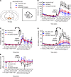
Figure 5. Amphetamine-induced rotations reveal differential DA-controlled behaviors following unilateral expression of DAT-WT and DAT-K619N.
The effect of HA-DAT-WT and HA-DAT-K619N on striatal DA homeostasis was compared by unilateral expression and open-field assessment of amphetamine-induced rotations. (A) TH-Cre mice were injected unilaterally in substantia nigra with AAV encoding either HA-DAT-WT, HA-DAT-K619N, or mCherry as a control plasmid. Amphetamine-induced rotations were evaluated in the open-field setup 3 weeks after injections. (B) Distance travelled in open-field chambers 90 minutes before and after amphetamine injections (5 mg/kg, i.p.). Mice injected with HA-DAT-WT differed from HA-DAT-K619N–injected mice by driving enhanced activity relative to both mCherry-injected and HA-DAT-K619N–injected mice (repeated measures 2-way ANOVA followed by Holm-Šídák multiple-comparison test, n = 7–9 mice). (C) Evaluation of ipsilateral rotations showed no difference between mice injected with HA-DAT-WT, HA-DAT-K619N, or mCherry upon amphetamine exposure (P > 0.05, repeated measures 2-way ANOVA). (D) The number of contralateral rotations after amphetamine treatment was enhanced only in mice expressing HA-DAT-WT and not in HA-DAT-K619N–expressing mice, supporting the differential effects of HA-DAT-K619N and HA-DAT-WT in vivo. (E) Rotational laterality assessed as ipsilateral-contralateral rotations before and after amphetamine injections. Only HA-DAT-WT–injected mice displayed lateralized rotational behavior (2-way ANOVA followed by Holm-Šídák multiple-comparison test, n = 7–9 mice). In panels B, D, and E, plus signs mark statistical differences between HA-DAT-WT and HA-DAT-K619N, and asterisks mark statistical differences between HA-DAT-WT and mCherry. No differences were found between HA-DAT-K619N and mCherry. ****/++++P < 0.0001, ***/+++P < 0.001, **/++P < 0.01, */+P < 0.05). All data are shown as mean ± SEM.
Figure 6
Figure 6. DAT-K619N exerts a dominant-negative effect on DAT-WT.
(A) Evaluation of dominant-negative effects of DAT-K619N in vitro. DA uptake was measured in HEK293 cells, cotransfected with equal amounts (1.5 μg) of DAT-K619N and DAT-WT, and compared with cells transfected only with DAT-WT (1.5 μg + 1.5 μg empty vector), DAT-K619N (1.5 μg + 1.5 μg empty vector), or with 3 μg of DAT-WT as a control. Each experiment was performed in triplicate and normalized to Vmax of DAT-WT (1.5 μg + 1.5 μg empty vector). The Vmax of DAT-K619N/DAT-WT cotransfected cells (1.5 μg + 1.5 μg) was reduced relative to DAT-WT (1.5 μg + 1.5 μg empty vector) to a level similar to DAT-K619N alone (*P < 0.016, 1-sample 2-tailed t test, Bonferroni-adjusted significance level [= 0.05/3] n = 4–7) indicating dominant-negative actions of hDAT-K619N. (B) Evaluation of dominant-negative effects in vivo performed by overexpressing HA-hDAT-WT or HA-hDAT-K619N selectively in dopaminergic neurons, using bilateral midbrain AAV injections in TH-Cre mice, and performing [3H]-DA uptake on striatal synaptosomes. Injections with AAV encoding mCherry was used for comparison with endogenous DA uptake levels. Uptake curves are average curves of 4 experiments each performed in triplicate. Quantification of Vmax normalized to mCherry showed HA-hDAT-K619N but not HA-hDAT-WT reduced DA uptake below endogenous uptake capacity (*P < 0.005, 1-sample 2-tailed t test, Bonferroni-adjusted significance level: **P < 0.01/2; n = 4. (C) Western blot analysis of striatal synaptosomal preparations. Representative blots for total DAT, HA-DAT, and TH are shown. Specific detection of HA-hDAT-WT and HA-hDAT-K619N using anti-HA antibody confirmed both constructs were trafficked to striatal terminals. (DF) Quantification of relative expression levels revealed a reduction in both DAT (E) and TH (F) in HA-hDAT-K619N–injected mice (*P < 0.025, 1-sample 2-tailed t test, Bonferroni-adjusted significance level [= 0.05/2], n = 4); HA-DAT levels did not differ significantly between HA-DAT-WT and HA-DAT-K619N–injected mice (P = 0.21, 1-sample 2-tailed t test, n = 4).

References

    1. Iversen SD, Iversen LL. Dopamine: 50 years in perspective. Trends Neurosci. 2007;30(5):188–193. doi: 10.1016/j.tins.2007.03.002. - DOI - PubMed
    1. Berke JD. What does dopamine mean? Nat Neurosci. 2018;21(6):787–793. doi: 10.1038/s41593-018-0152-y. - DOI - PMC - PubMed
    1. Del Campo N, et al. The roles of dopamine and noradrenaline in the pathophysiology and treatment of attention-deficit/hyperactivity disorder. Biol Psychiatry. 2011;69(12):e145–e157. doi: 10.1016/j.biopsych.2011.02.036. - DOI - PubMed
    1. Dichter GS, et al. Reward circuitry dysfunction in psychiatric and neurodevelopmental disorders and genetic syndromes: animal models and clinical findings. J Neurodev Disord. 2012;4(1):19. doi: 10.1186/1866-1955-4-19. - DOI - PMC - PubMed
    1. Cousins DA, et al. The role of dopamine in bipolar disorder. Bipolar Disord. 2009;11(8):787–806. doi: 10.1111/j.1399-5618.2009.00760.x. - DOI - PubMed

Publication types

MeSH terms

Substances