. 2021 Aug 9;31(15):R938-R945.
doi: 10.1016/j.cub.2021.06.093.

Mucus

Affiliations

Mucus

Abigail McShane et al. Curr Biol. .

Abstract

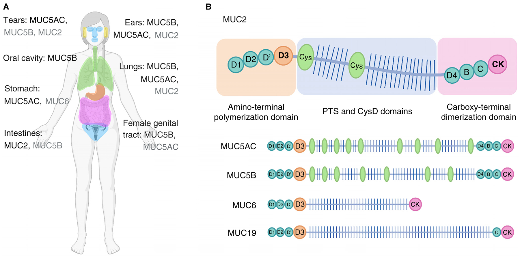
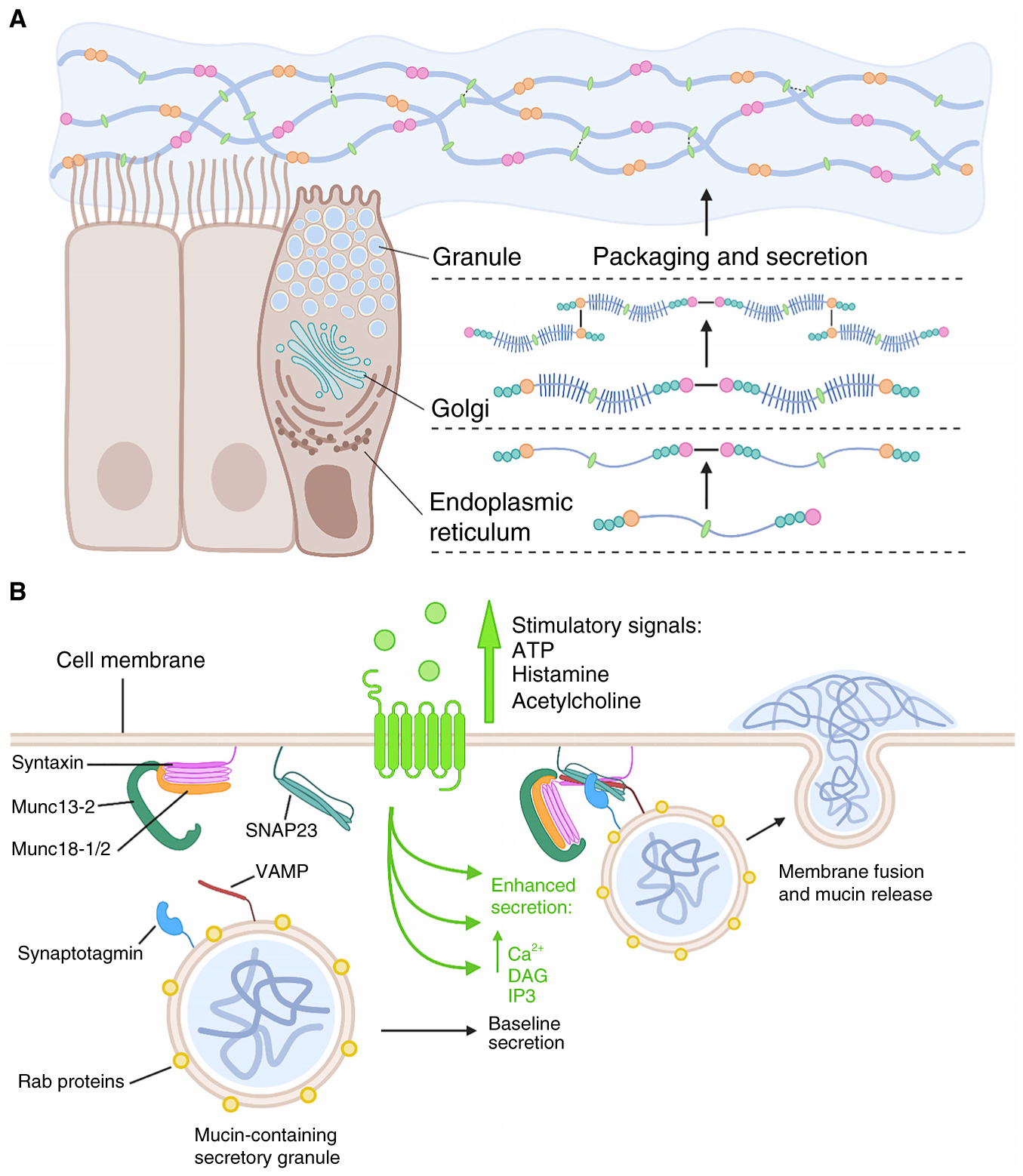
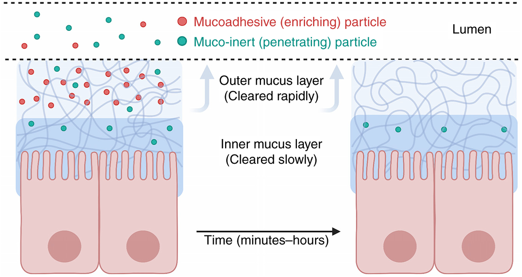
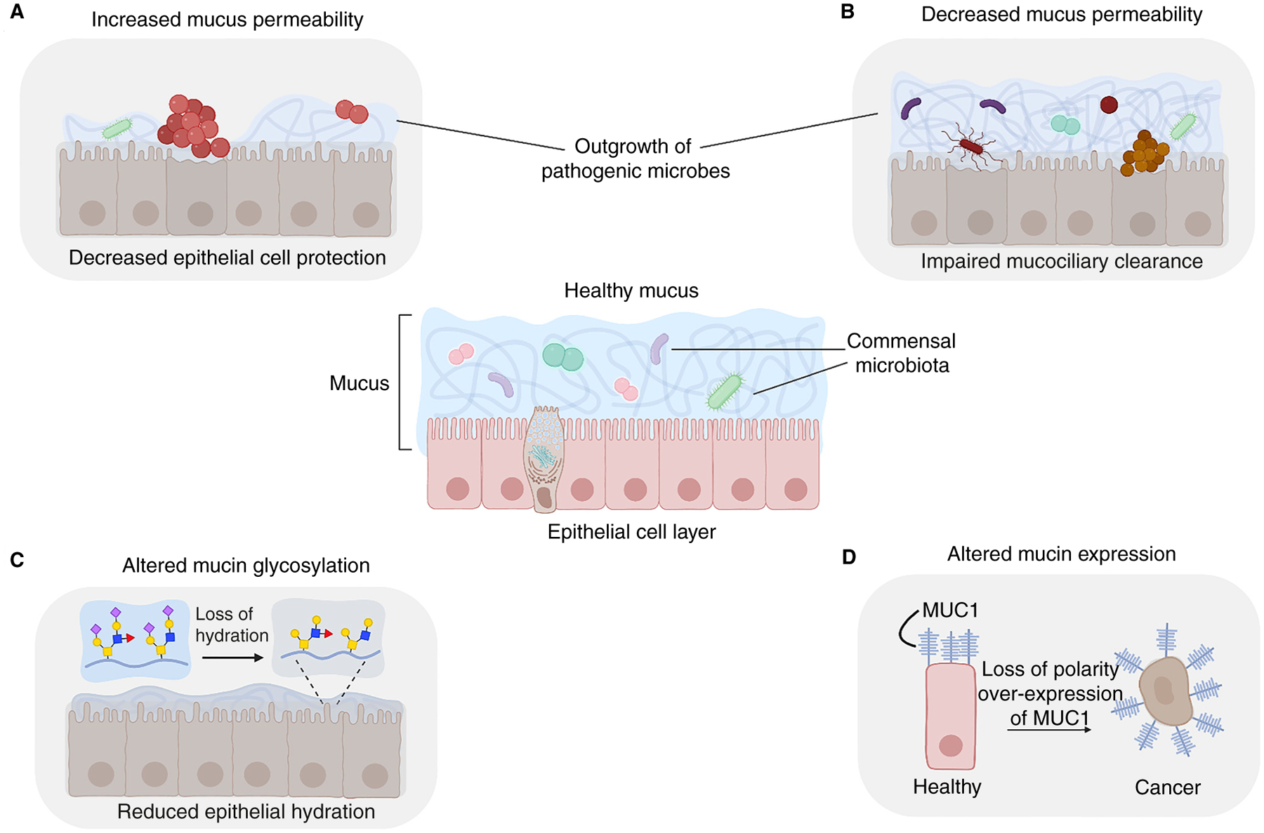
Mucus is a slimy hydrogel that lines the mucosal surfaces in our body, including the intestines, stomach, eyes, lungs and urogenital tract. This glycoprotein-rich network is truly the jack of all trades. As a barrier, it lubricates surfaces, protects our cells from physical stress, and selectively allows the passage of nutrients while clearing out pathogens and debris. As a home to our microbiota, it supports a level of microbial diversity that is unattainable with most culture methods. As a reservoir of complex carbohydrate structures called glycans, it plays critical roles in controlling cell adhesion and signaling, and it alters the behavior and spatial distribution of microbes. On top of all this, mucus regulates the passage of sperm during fertilization, heals wounds, helps us smell, and prevents the stomach from digesting itself, to name just a few of its functions. Given these impressive features, it is no wonder that mucus crosses boundaries of species and kingdoms - mucus gels are made by organisms ranging from the simplest metazoans to corals, snails, fish, and frogs. It is also no surprise that mucus is exploited in everyday applications, including foods, cosmetics, and other products relevant to medicine and industry.


Figures

Figure 1.
Figure 1.. Locations of secreted mucins in the body and their structural diversity.
(A) Mucus coats all non-keratinized wet epithelial surfaces. Various MUC genes are expressed throughout the body to form mucin gels that are adapted to various physiological environments. For tissues expressing multiple mucin types, the dominant mucins are listed in black, with non-dominant mucin types in grey. (B) Summary of the features of the gel-forming mucin, MUC2. The cartoon illustrates the domain organization of MUC2 (not drawn to scale), highlighting the vWF-like cysteine-rich domains at the amino and carboxy termini (B, C, and D domains; teal circles), the centrally located, highly O-glycosylated PTS domains (glycans shown as lines), and the internal cysteine-rich domains (CysD; green ovals). The amino- and carboxy-terminal domains are essential for polymer formation. Mucin polymers are stabilized by tail-to-tail disulfide linkages between carboxy-terminal CK domains (pink) and head-to-head disulfide linkages between amino-terminal vWF-like D3 domains (orange circles). (All figures created with BioRender.com.)
Figure 2.
Figure 2.. Mucins are synthesized, packaged and secreted from specialized cells to form the mucus gel.
(A) In brief, CK-domain-mediated dimerization of the mucin polypeptide occurs in the endoplasmic reticulum followed by O-glycan addition in the Golgi and then D3-domain-mediated multimerization. Assembled polymers are then compacted for storage in secretory granules via calcium- and pH-dependent mechanisms. After secretion, the polymers hydrate and expand to form a dynamic mucin network that is stabilized by a combination of non-covalent and covalent interactions; the relative contributions of these types of interactions are tissue-dependent, with intestinal mucus containing the highest level of covalent interactions stabilizing its structure. Colors and symbols are as defined in Figure 1. (B) Exocytosis of mucin granules is highly regulated. In the resting state (left), mucin-containing granules decorated with Rab proteins become tethered to the plasma membrane by interacting with Munc13-2 and other tethering proteins. Stimulatory signals (light green) binding to heptahelical receptors lead to the generation of second messengers diacylglycerol (DAG) and inositol triphosphate (IP3). DAG activates Munc13-2 and, together with Munc18, opens syntaxin, which then interacts with the other SNARE proteins SNAP23 and VAMP to form a four-helix-bundle SNARE complex (right). IP3 induces the release of Ca2+ from the ER that in turn activates the calcium sensor synaptotagmin, which promotes further coiling of the SNARE complex. This action drives the fusion of the mucin-containing granule with the plasma membrane and the subsequent release of mucins into the extracellular space. Both baseline and stimulated mucin-granule exocytosis are regulated, as they are dependent on extracellular signals and second messengers.
Figure 3.
Figure 3.. Many glycosyltransferases contribute to the generation of mucin-type O-glycan diversity.
Serine and threonine residues act as potential sites of O-glycosylation within PTS domains. At least 15 ppGalNAc transferases can accomplish the first step, initiation, in which a GalNAc is transferred to a serine or threonine residue. As the O-glycosylated mucin moves through later compartments of the Golgi apparatus, glycan residues are further extended by at least 30 different additional glycosyltransferases with distinct donor and acceptor sugars using both α and β linkages. This combinatorial approach generates a plethora of diverse glycan structures based on eight core scaffolds, with cores 1–4 being the most common. Representative glycan structures present on human gastric mucin are shown.
Figure 4.
Figure 4.. Molecular properties influence enrichment and longevity within mucus layers.
Particles with concentrated regions of positive charge or hydrophobicity can bind to and become enriched in mucus layers (left, red dots). However, binding to mucus leads to faster clearance from the body due to sloughing off of the outer mucus layer. Particles that do not bind to mucus do not become enriched in mucus layers but are able to penetrate deeper and are not cleared as quickly from the body (right, blue dots). Both mucoadhesive and muco-inert particles present advantages and tradeoffs in terms of drug design and delivery. (This figure was adapted from Lai et al. (2009).)
Figure 5.
Figure 5.. Healthy mucus protects against barrier dysfunction and disease.
Healthy mucus (center) protects epithelia by acting as a dynamic barrier that provides hydration, regulates diffusion and accommodates commensal microbes. Changes in mucin expression and identity are associated with disease. (A) Increased mucus permeability in various tissues potentially allows the outgrowth of pathogenic microbes leading to intra-amniotic infections and stomach ulcers. (B) Decreased mucus permeability impairs mucociliary clearance, leading to increased microbial colonization, as observed in cystic fibrosis and chronic obstructive pulmonary disease. (C) Altered mucin glycosylation is associated with various diseases, including Sjögren’s syndrome, cancer, and cystic fibrosis. Loss of sialylated glycans in Sjögren’s syndrome reduces the hydrophilicity of mucin glycans, causing dry mouth. (D) Altered mucin expression can lead to cancer. Membrane-associated MUC1 promotes the survival of epithelial cells, but MUC1 overexpression can lead to aberrant regulation of growth factor signaling.

References

    1. Bakshani CR, Morales-Garcia AL, Althaus M, Wilcox MD, Pearson JP, Bythell JC, and Burgess JG (2018). Evolutionary conservation of the antimicrobial function of mucus: a first defence against infection. NPJ Biofilms Microbiomes 4, 14. - PMC - PubMed
    1. Bansil R, and Turner BS (2018). The biology of mucus: Composition, synthesis and organization. Adv. Drug Deliv. Rev 124, 3–15. - PubMed
    1. Bennett EP, Mandel U, Clausen H, Gerken TA, Fritz TA, and Tabak LA (2012). Control of mucin-type O-glycosylation: A classification of the polypeptide GalNAc-transferase gene family. Glycobiology 22, 736–756. - PMC - PubMed
    1. Boucher RC (2019). Muco-obstructive lung diseases. N. Engl. J. Med 380, 1941–1953. - PubMed
    1. Campbell L, Hepworth MR, Whittingham-Dowd J, Thompson S, Bancroft AJ, Hayes KS, Shaw TN, Dickey BF, Flamar A-L, Artiset E, et al. (2019). ILC2s mediate systemic innate protection by priming mucus production at distal mucosal sites. J. Exp. Med 216, 2714–2723. - PMC - PubMed