Dual-Energy Computed Tomography Compared to Lung Perfusion Scintigraphy to Assess Pulmonary Perfusion in Patients Screened for Endoscopic Lung Volume Reduction
- PMID: 34375973
- PMCID: PMC8743912
- DOI: 10.1159/000517598
Dual-Energy Computed Tomography Compared to Lung Perfusion Scintigraphy to Assess Pulmonary Perfusion in Patients Screened for Endoscopic Lung Volume Reduction
Abstract
Background: Endoscopic lung volume reduction (ELVR) using one-way endobronchial valves is a technique to reduce hyperinflation in patients with severe emphysema by inducing collapse of a severely destroyed pulmonary lobe. Patient selection is mainly based on evaluation of emphysema severity on high-resolution computed tomography and evaluation of lung perfusion with perfusion scintigraphy. Dual-energy contrast-enhanced CT scans may be useful for perfusion assessment in emphysema but has not been compared against perfusion scintigraphy.
Aims: The aim of the study was to compare perfusion distribution assessed with dual-energy contrast-enhanced computed tomography and perfusion scintigraphy.
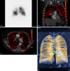
Material and methods: Forty consecutive patients with severe emphysema, who were screened for ELVR, were included. Perfusion was assessed with 99mTc perfusion scintigraphy and using the iodine map calculated from the dual-energy contrast-enhanced CT scans. Perfusion distribution was calculated as usually for the upper, middle, and lower thirds of both lungs with the planar technique and the iodine overlay.
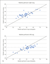
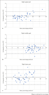
Results: Perfusion distribution between the right and left lung showed good correlation (r = 0.8). The limits of agreement of the mean absolute difference in percentage perfusion per region of interest were 0.75-5.6%. The upper lobes showed more severe perfusion reduction than the lower lobes. Mean difference in measured pulmonary perfusion ranged from -2.8% to 2.3%. Lower limit of agreement ranged from -8.9% to 4.6% and upper limit was 3.3-10.0%.
Conclusion: Quantification of perfusion distribution using planar 99mTc perfusion scintigraphy and iodine overlays calculated from dual-energy contrast-enhanced CTs correlates well with acceptable variability.
Keywords: Bronchoscopic lung volume reduction; Computed tomography, lung; Dual-energy computed tomography; Emphysema; Perfusion scan; Scintigraphy.
© 2021 The Author(s) Published by S. Karger AG, Basel.
Conflict of interest statement
The authors have no conflicts of interest to disclose.
Figures


References
-
- Herth FJF, Slebos DJ, Criner GJ, Valipour A, Sciurba F, Shah PL. Endoscopic lung volume reduction: an expert panel recommendationUpdate 2019. Respiration. 2019;97((6)):548–57. - PubMed
-
- Valipour A, Slebos DJ, Herth F, Darwiche K, Wagner M, Ficker JH, et al. Endobronchial valve therapy in patients with homogeneous emphysema. Results from the IMPACT study. Am J Respir Crit Care Med. 2016;194((9)):1073–82. - PubMed
-
- Koster TD, van Rikxoort EM, Huebner RH, Doellinger F, Klooster K, Charbonnier JP, et al. Predicting lung volume reduction after endobronchial valve therapy is maximized using a combination of diagnostic tools. Respiration. 2016;92((3)):150–7. - PubMed
Publication types
MeSH terms
Substances
LinkOut - more resources
Full Text Sources
Medical
Research Materials

