

Endoscopic and fluoroscopic-guided closure of the eustachian tube using a biliary cytology brush and liquid embolic agent for a persistent CSF leak after schwannoma resection
- PMID: 34376411
- PMCID: PMC8356153
- DOI: 10.1136/bcr-2021-241861
Endoscopic and fluoroscopic-guided closure of the eustachian tube using a biliary cytology brush and liquid embolic agent for a persistent CSF leak after schwannoma resection
Abstract
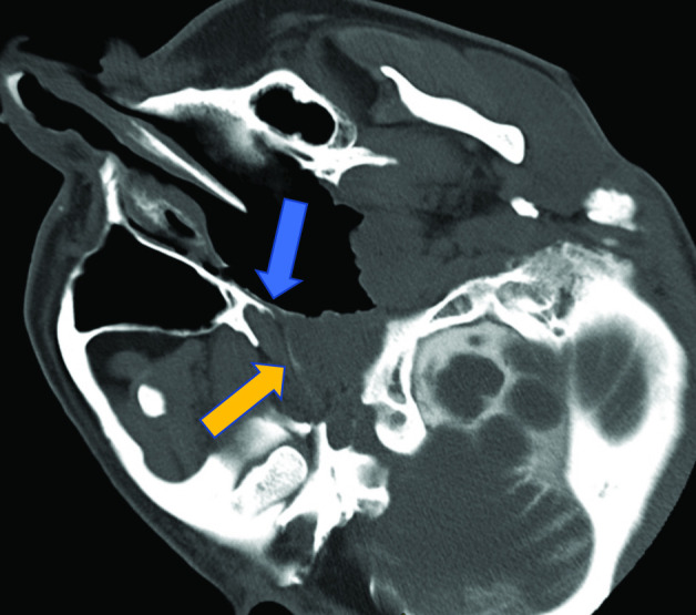
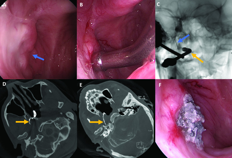
Vestibular schwannoma is a known cause of progressive sensorineural hearing loss. Treatment options include observation, radiation therapy and surgical resection. Cerebrospinal fluid (CSF) fistula is a known postsurgical complication that can lead to CSF otorrhoea, rhinorrhoea or CSF leakage from the surgical wound. We present a case report of a patient who underwent vestibular schwannoma resection and postoperatively developed CSF rhinorrhoea, which was refractory to multiple attempts at surgical repair. This was successfully treated under endoscopic and fluoroscopic guidance using a biliary cytology brush to disrupt the surface of the eustachian tube followed by injection of n-Butyl cyanoacrylate.
Keywords: ear; interventional radiology; nose and throat/otolaryngology.
© BMJ Publishing Group Limited 2021. No commercial re-use. See rights and permissions. Published by BMJ.
Conflict of interest statement
Competing interests: None declared.
Figures
Similar articles
-
Transnasal Fluoroscopic-Guided Eustachian Tube Obliteration With a Liquid Embolic Agent for a Recurrent Cerebrospinal Fluid Leak After Translabyrinthine Schwannoma Surgery: A Case Series.Otol Neurotol. 2025 Apr 1;46(4):e125-e129. doi: 10.1097/MAO.0000000000004428. Epub 2025 Feb 4. Otol Neurotol. 2025. PMID: 39965222
-
Endoscopic closure of the eustachian tube for repair of cerebrospinal fluid leak.Am J Otol. 1996 May;17(3):470-2. Am J Otol. 1996. PMID: 8817027
-
Managing cerebrospinal fluid rhinorrhea after lateral skull base surgery via endoscopic endonasal eustachian tube closure.Am J Rhinol Allergy. 2015 May-Jun;29(3):207-10. doi: 10.2500/ajra.2015.29.4146. Am J Rhinol Allergy. 2015. PMID: 25975252
-
Minimally invasive endoscopic repair of refractory lateral skull base cerebrospinal fluid rhinorrhea: case report and review of the literature.Neurosurg Focus. 2018 Mar;44(3):E8. doi: 10.3171/2017.12.FOCUS17664. Neurosurg Focus. 2018. PMID: 29490552 Review.
-
Management options for cerebrospinal fluid leak after vestibular schwannoma surgery and introduction of an innovative treatment.Otol Neurotol. 2004 Jul;25(4):580-6. doi: 10.1097/00129492-200407000-00027. Otol Neurotol. 2004. PMID: 15241238 Review.
References
Publication types
MeSH terms
LinkOut - more resources
Full Text Sources