. 2021 Apr;15(s1):10-25.
doi: 10.14444/8052. Epub 2021 Apr 21.

Pathomechanism and Biomechanics of Degenerative Disc Disease: Features of Healthy and Degenerated Discs

Affiliations

Pathomechanism and Biomechanics of Degenerative Disc Disease: Features of Healthy and Degenerated Discs

Sertac Kirnaz et al. Int J Spine Surg. 2021 Apr.

Abstract

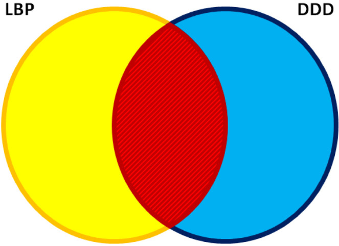
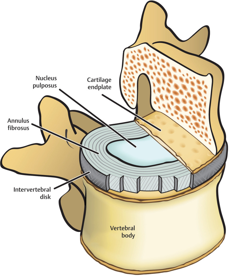
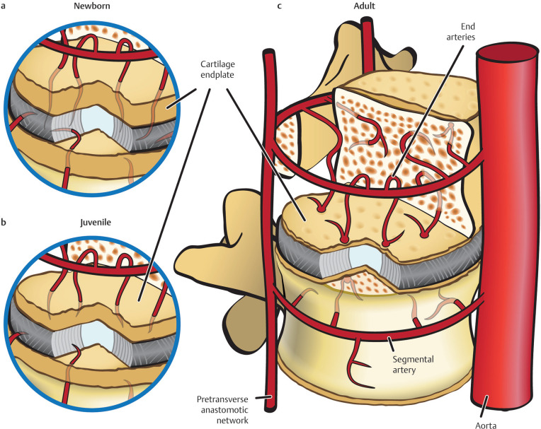
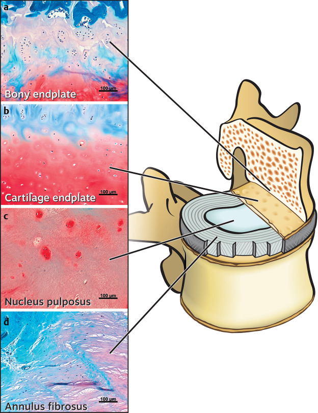
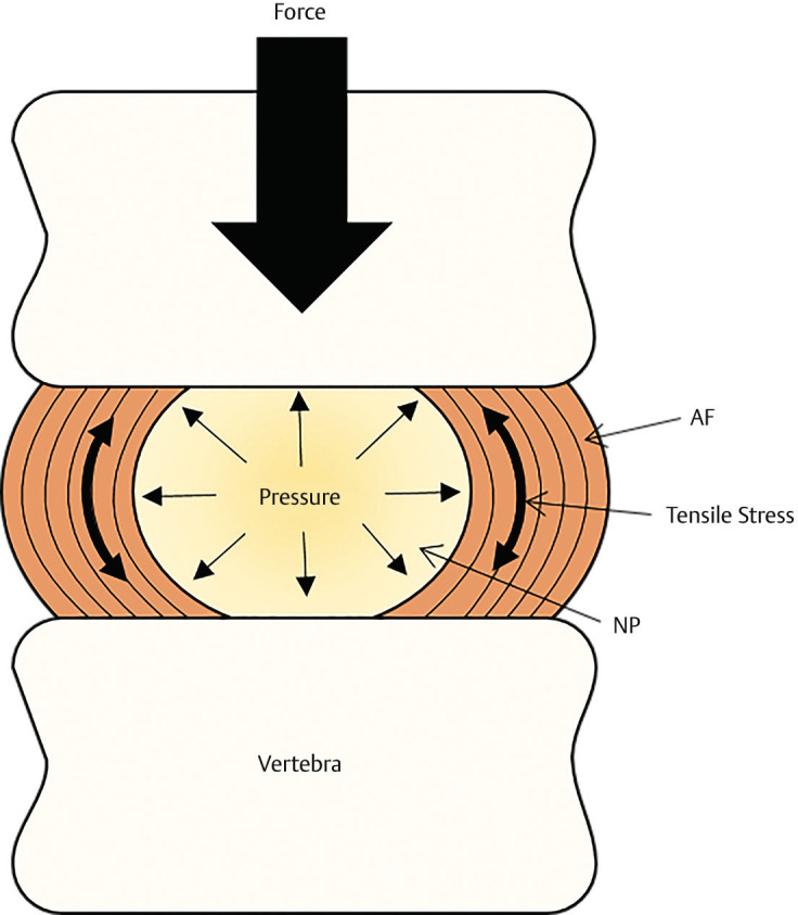
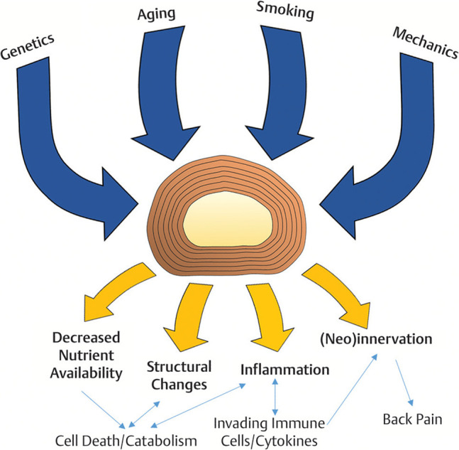
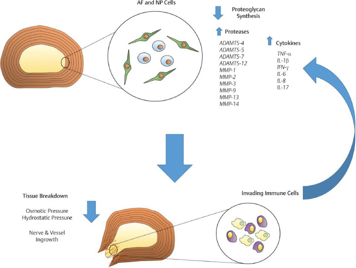
The human intervertebral disc (IVD) is a complex organ composed of fibrous and cartilaginous connective tissues, and it serves as a boundary between 2 adjacent vertebrae. It provides a limited range of motion in the torso as well as stability during axial compression, rotation, and bending. Adult IVDs have poor innate healing potential due to low vascularity and cellularity. Degenerative disc disease (DDD) generally arises from the disruption of the homeostasis maintained by the structures of the IVD, and genetic and environmental factors can accelerate the progression of the disease. Impaired cell metabolism due to pH alteration and poor nutrition may lead to autophagy and disruption of the homeostasis within the IVD and thus plays a key role in DDD etiology. To develop regenerative therapies for degenerated discs, future studies must aim to restore both anatomical and biomechanical properties of the IVDs. The objective of this review is to give a detailed overview about anatomical, radiological, and biomechanical features of the IVDs as well as discuss the structural and functional changes that occur during the degeneration process.

Keywords: back pain; biomechanics; degenerative disc disease; intervertebral disc; low back pain; lumbar disc herniation; pathophysiology.


Conflict of interest statement

Disclosures and COI: Roger Härtl is a consultant for Ulrich, Brainlab, DePuy-Synthes, and he has royalties from Zimmer. The authors received no funding for this study.

Figures

Figure 1
Figure 1
Low back pain (LBP) is a clinical entity, and degenerative disc disease (DDD) is a radiographic-anatomical finding. There are many reasons why patients can present with LBP. Conversely, DDD is frequently found in imaging studies of asymptomatic patients. However, there is an overlap, and patients with LBP can present with imaging findings consistent with DDD. The challenge for the clinician then is to establish whether there is a causative relationship.
Figure 2
Figure 2
Anatomical composition of the human intervertebral disc. Centrally located nucleus pulposus and concentric organization of annulus fibrosus lamellae surrounding the periphery. Superiorly and inferiorly cover by cartilaginous endplate. Printed with permission from Biological Approaches to Spinal Disc Repair and Regeneration for Clinicians.
Figure 3
Figure 3
(a) Neonate's vertebral body and disc vascular network; blood supply can extend into the innermost regions of the annulus fibrosis (AF). (b) Vessels retract further from the disc to the outer region of the AF during adolescence. (c) Vessels are regressed further away from the AF and fix themselves within and surrounding the end plate and connective tissues in adult spines. Printed with permission from Biological Approaches to Spinal Disc Repair and Regeneration for Clinicians.
Figure 4
Figure 4
Histology images of human intervertebral disc. (a) and (b) Bony and cartilaginous endplates of a young healthy patient. The bony endplate is distinct and contains hypertrophic cartilage. (c) and (d) The nucleus pulpous (NP) and annulus fibrosus (AF). Histology stains were Safranin-O, fast green FCF, and Weigert's hematoxylin. Printed with permission from Biological Approaches to Spinal Disc Repair and Regeneration for Clinicians.
Figure 5
Figure 5
The intervertebral disc (IVD) undergoes load bearing, bending, flexing, and torsion while under mechanical stressors. The annulus fibrosus (AF) and the nucleus pulposus (NP) form the IVD within the intervertebral space to fill the joint and maintain disc height. When the IVD is undergoes the previously mentioned physical stress, then the NP reacts by resisting the downward force against it by pressing vertically back into the compression and radially into the AF. The pressure from the NP generates tensile stress on the AF in the direction of the organization of the fibers within the AF. Printed with permission from Biological Approaches to Spinal Disc Repair and Regeneration for Clinicians.
Figure 6
Figure 6
The sinuvertebral nerve innervates the disc. In a healthy adult disc, the nerve endings cannot reach into the innermost layers of the annulus fibrosus (AF). Printed with permission from Biological Approaches to Spinal Disc Repair and Regeneration for Clinicians.
Figure 7
Figure 7
Degenerative disc disease (DDD) in a cascading multifactorial process involving the interaction of risk factors and pathophysiology. Printed with permission from Biological Approaches to Spinal Disc Repair and Regeneration for Clinicians.
Figure 8
Figure 8
The physiological alterations to the disc are controlled directly by cells within the disc, which are caught in a closed degenerative cycle. These cells increase production of cytokines and proteases while decreasing production of proteoglycans, both of which are essential to the retaining the disc's height as well as maintaining the basic physical function of the intervertebral disc. The increase of proteases expedites the tissue degeneration process. The proteases also alter the extracellular environment, which incurs catabolic reaction and inflammation. This process results in the activation of nearby immune cells that exacerbates the inflammatory processes by continuing to increase cytokine production. The increase in cytokines enhances neovascularization and neoinnervation in the disc. Printed with permission from Biological Approaches to Spinal Disc Repair and Regeneration for Clinicians.
Figure 9
Figure 9
T2-weighted sagittal magnetic resonance images of three different patients. Roman numerals present the Pfirrmann grades. (Left) A female adolescent patient with healthy discs, (center) a young adult female patient with mild disc degeneration, and (right) a senior male patient with advanced disc degeneration. Printed with permission from Biological Approaches to Spinal Disc Repair and Regeneration for Clinicians.

Similar articles

Cited by

References

    1. Hoy D, March L, Brooks P, et al. The global burden of low back pain: estimates from the Global Burden of Disease 2010 study. Ann Rheum Dis. 2014;73(6):968–974. - PubMed
    1. Hoy D, Brooks P, Blyth F, Buchbinder R. The epidemiology of low back pain. Best Prac Res Cl Rh. 2010;24(6):769–781. - PubMed
    1. Hooten WM, Cohen SP. Evaluation and treatment of low back pain: a clinically focused review for primary care specialists. Mayo Clin Proc. 2015;90(12):1699–1718. - PubMed
    1. Anderson DG, Tannoury C. Molecular pathogenic factors in symptomatic disc degeneration. Spine J. 2005;5(suppl 6):260s–266s. - PubMed
    1. Zhang YG, Guo TM, Guo X, Wu SX. Clinical diagnosis for discogenic low back pain. Int J Biol Sci. 2009;5(7):647–658. - PMC - PubMed