. 2021 Aug 31;30(4):285-293.
doi: 10.5607/en21022.

Region-Specific Differences in the Apoe4-dependent Response to Focal Brain Injury

Affiliations

Region-Specific Differences in the Apoe4-dependent Response to Focal Brain Injury

Sung Eun Lee et al. Exp Neurobiol. .

Abstract

Apolipoprotein E (apoE) plays a role in various physiological functions including lipid transport, synaptic plasticity, and immune modulation. Epidemiological studies suggest that the apoE4 allele increases the risk of post-traumatic sequelae. This study was performed to investigate regionspecific effects of the apoE4 isoform on post-traumatic neurodegeneration. Two focal brain injuries were introduced separately in the motor cortex and hippocampus of apoE4 knock-in, apoE3 knock-in, apoE knockout, and wild-type (WT) mice. Western blotting showed that the expression levels of pre-synaptic and post-synaptic markers at the recovery stage were lower in the hippocampal injury core in apoE4 mice, compared with apoE3 and WT mice. Fast glial activation (determined by immunohistochemistry with glial fibrillary acidic protein, ionized calcium binding adaptor molecule 1, and cluster of differentiation 45 antibodies) was characteristic of apoE4 mice with hippocampal injury penumbra. apoE4-specific changes were not observed after cortical injury. The intensity of microglial activation in the hippocampus was inversely correlated with the volume of injury reduction on sequential magnetic resonance imaging examinations, when validated using matched samples. These findings indicate that the effects of the interaction between apoE4 and focal brain damage are specific to the hippocampus. Manipulation of inflammatory cell responses could be beneficial for reducing post-traumatic hippocampal neurodegeneration in apoE4 carriers.

Keywords: Apolipoprotein E4; Brain injuries; Hippocampus; Inflammation; Neurodegeneration; Neuroglia.


Figures

Fig. 1
Fig. 1
Experimental flow. MRIs were taken on days 1 and 15 post-injury. Tissue was prepared on days 3 (n=19) or 15 (n=36) after ATP injection for immunohistochemistry and western blot. IHC, immunohistochemistry; KI, knock-in; WB, western blot.
Fig. 2
Fig. 2
Focal brain injury and its recovery on brain MRI. (A) MRI on day 1 after ATP injection showed well-demarcated focal injuries in the motor cortex (M1) and hippocampus (CA1) as T2-hyperintense signals (arrowheads). The volumes of injured tissue were comparable among apoE genotypes (all p>0.05). (B) Damaged regions were shrunken and magnetic resonance signals exhibited lower intensity on day 15. The volumes of remaining injuries were similar in all groups (all p>0.05). (C) Damage reduction on day 15, compared with day 1, did not differ in the cortex and hippocampus across all groups (all p>0.05). Bar graphs show means±standard errors of the mean. Box plots show medians and interquartile ranges, while whiskers represent maximum and minimum values. *p<0.05, significant on one-way analysis of variance with post hoc Tukey’s test. 3KI, apoE3 knock-in; 4KI, apoE4 knock-in; KO, knockout; vol, volume; wt, wild-type.
Fig. 3
Fig. 3
Western blotting analysis of the injury core on day 15. Western blotting and densitometric analyses showed comparable NeuN expression levels in all groups in both the motor cortex (M1) and hippocampus (CA1) (p>0.05). The expression of SYP was significantly decreased upon cortical injury of 3KI and 4KI with respect to wild-type mice (p=0.029), whereas PSD-95 levels were equivalent. Both SYP and PSD-95 expression levels in the hippocampus showed isoform-dependent changes: significant reductions in 4KI and KO mice, compared with 3KI and wild-type mice (p=0.017 and p=0.041, respectively). Bar graphs show means±standard errors of the mean. *p<0.05, significant on Kruskal–Wallis test and subsequent post hoc analyses using the Mann–Whitney test with multiple comparison correction by means of FDR<0.1. 3KI, apoE3 knock-in; 4KI, apoE4 knock-in; ct, control mice without injury; IC, repeatedly loaded sample to compare two blots; KO, knockout; NeuN, neuronal nuclear protein; SYP, synaptophysin; PSD-95, postsynaptic density-95; wt, wild-type.
Fig. 4
Fig. 4
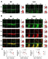
Immunohistochemical assessment of glial markers and correlation analysis. (A) GFAP immunohistochemical assessment showed higher astrocytic activation upon hippocampal injury in 4KI mice, compared with 3KI mice on day 3 (p=0.042) and compared with KO mice on day 15 (p=0.048). (B) Iba-1+ and Iba-1+/CD45+ signals were significantly lower in 3KI mice than in wild-type mice upon cortical injury on day 3 (p=0.016 and p=0.011, respectively), and the Iba-1+ signal was higher in 4KI mice than in KO mice on day 15 (p=0.038). Iba-1, CD45, and Iba-1/CD45 immunoreactivities in the hippocampus were all higher in 4KI mice than in 3KI mice on day 3 (p=0.009, p=0.019, and p=0.009, respectively). These markers further elevated on day 15, although the group differences disappeared. (C) A significant inverse correlation between injury volume reduction on MRI and Iba-1 immunoreactivity (r=-0.434, p=0.049), and a tendency for inverse correlation between MRI marker and Iba-1+/CD45+ (r=-0.430, p=0.052), were identified in correlation analysis. The asterisks (*) in figures indicate the side of injury core. The dotted lines show the border of injury core. Bar graphs show mean±standard errors of the mean. Scale bar, 100 μm. *p<0.05 and **p<0.01, significant on one-way analysis of variance with post hoc Tukey’s test (A, B) and Pearson’s correlation analysis (C). γ, correlation coefficients; 3KI, apoE3 knock-in; 4KI, apoE4 knock-in; GFAP, glial fibrillary acidic protein; KO, knockout; wt, wild-type.

Similar articles

Cited by

References

    1. Wang H, Eckel RH. What are lipoproteins doing in the brain? Trends Endocrinol Metab. 2014;25:8–14. doi: 10.1016/j.tem.2013.10.003. - DOI - PMC - PubMed
    1. Hu J, Liu CC, Chen XF, Zhang YW, Xu H, Bu G. Opposing effects of viral mediated brain expression of apolipoprotein E2 (apoE2) and apoE4 on apoE lipidation and Aβ metabolism in apoE4-targeted replacement mice. Mol Neurodegener. 2015;10:6. doi: 10.1186/s13024-015-0001-3. - DOI - PMC - PubMed
    1. Teter B. ApoE-dependent plasticity in Alzheimer's disease. J Mol Neurosci. 2004;23:167–179. doi: 10.1385/JMN:23:3:167. - DOI - PubMed
    1. Crawford FC, Vanderploeg RD, Freeman MJ, Singh S, Waisman M, Michaels L, Abdullah L, Warden D, Lipsky R, Salazar A, Mullan MJ. APOE genotype influences acquisition and recall following traumatic brain injury. Neurology. 2002;58:1115–1118. doi: 10.1212/WNL.58.7.1115. - DOI - PubMed
    1. Kassam I, Gagnon F, Cusimano MD. Association of the APOE-ε4 allele with outcome of traumatic brain injury in children and youth: a meta-analysis and meta-regression. J Neurol Neurosurg Psychiatry. 2016;87:433–440. doi: 10.1136/jnnp-2015-310500. - DOI - PubMed