. 2021 Aug 10;8(1):191.
doi: 10.1038/s41438-021-00625-9.

The transcription factor CmLEC1 positively regulates the seed-setting rate in hybridization breeding of chrysanthemum

Affiliations

The transcription factor CmLEC1 positively regulates the seed-setting rate in hybridization breeding of chrysanthemum

Sujuan Xu et al. Hortic Res. .

Abstract

Distant hybridization is widely used to develop crop cultivars, whereas the hybridization process of embryo abortion often severely reduces the sought-after breeding effect. The LEAFY COTYLEDON1 (LEC1) gene has been extensively investigated as a central regulator of seed development, but it is far less studied in crop hybridization breeding. Here we investigated the function and regulation mechanism of CmLEC1 from Chrysanthemum morifolium during its seed development in chrysanthemum hybridization. CmLEC1 encodes a nucleic protein and is specifically expressed in embryos. CmLEC1's overexpression significantly promoted the seed-setting rate of the cross, while the rate was significantly decreased in the amiR-CmLEC1 transgenic chrysanthemum. The RNA-Seq analysis of the developing hybrid embryos revealed that regulatory genes involved in seed development, namely, CmLEA (late embryogenesis abundant protein), CmOLE (oleosin), CmSSP (seed storage protein), and CmEM (embryonic protein), were upregulated in the OE (overexpressing) lines but downregulated in the amiR lines vs. wild-type lines. Future analysis demonstrated that CmLEC1 directly activated CmLEA expression and interacted with CmC3H, and this CmLEC1-CmC3H interaction could enhance the transactivation ability of CmLEC1 for the expression of CmLEA. Further, CmLEC1 was able to induce several other key genes related to embryo development. Taken together, our results show that CmLEC1 plays a positive role in the hybrid embryo development of chrysanthemum plants, which might involve activating CmLEA's expression and interacting with CmC3H. This may be a new pathway in the LEC1 regulatory network to promote seed development, one perhaps leading to a novel strategy to not only overcome embryo abortion during crop breeding but also increase the seed yield.


Conflict of interest statement

The authors declare no competing interests.

Figures

Fig. 1
Fig. 1. Isolation and sequence analysis of the CmLEC1 gene.
a Amino acid sequence alignment of CmLEC1 and plant LEC1 proteins, whose sequence features include a NF-YB/HAP3 domain. b Phylogenetic analysis of plants’ amino acid sequences of LEC1. c CmLEC1 is localized to the nucleus, based on transient expression profiles of CmLEC1 in N. benthamiana leaves. The co-expressed 35S::D53-RFP construct indicated the localization of nuclei. Scale bars = 5 μm. d CmLEC1 is specifically expressed in chrysanthemum embryos. NE12 normal embryos at 12 days after pollination, NE18 normal embryos at 18 days after pollination, AE18 abnormal embryos at 18 days after pollination. Error bars represent ±SD
Fig. 2
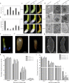
Fig. 2. Phenotype analysis of OE-CmLEC1 and amiR-CmLEC1 chrysanthemum transgenic lines.
a Relative expression level of CmLEC1 in OE-CmLEC1 plants. b Relative expression level of CmLEC1 in amiR-CmLEC1 plants. c Morphological features of the NE12, NE18, and NE25 in chrysanthemum ovaries of ♀OE-CmLEC1-C.m.×♂C.n. and ♀amiR-CmLEC1-C.m.×♂C.n. crosses. Scale bars = 1 mm. 12 DAP 12 days after pollination, 18 DAP 18 days after pollination, 25 DAP 25 days after pollination. d Transmission electron microscopy of NE12, NE18, and NE25 in chrysanthemum embryos of the ♀OE-CmLEC1-C.m.×♂C.n. and ♀amiR-CmLEC1-C.m.×♂C.n. crosses. Scale bar = 2 μm. Nu nucleolus, V vacuole, M mitochondria, N nucleus, ER endoplasmic reticulum, P plastid, CW cell wall. e Anatomical view of a chrysanthemum ovary. Scale bar = 0.978 mm. f Anatomical features of normal and abortive chrysanthemum ovaries. Scale bar = 500 μm. g Morphological characteristics of chrysanthemum ovaries. Scale bar = 20 μm. h Proportion of normal ovaries at different times after pollination in the ♀OE-CmLEC1-C.m.×♂C.n. cross. i Proportion of normal ovaries at different times after pollination in the ♀amiR-CmLEC1-C.m.×♂C.n. cross
Fig. 3
Fig. 3. RNA-Seq analysis of plants with an altered CmLEC1.
a Venn diagram of the number of genes in OE, amiR, and WT (wild-type) plants obtained by RNA-Seq. b Number of differentially expressed genes (DEGs) between OE, amiR, and WT obtained by RNA-Seq. c KEGG analysis; on the x-axis is the enrichment ratio, and on the y-axis are the KEGG pathways, for which a bubble’s size indicates the number of genes annotated to a certain KEGG pathway. d Heat map of the DEGs based on the RNA-Seq analysis of OE and amiR. The color scale indicates the scale of each gene expression level (log2FPKM). Rectangles in red denote genes’ upregulation, those in blue their downregulation. e Identification of the genes from DEGs related to seed development by qRT-PCR. The values are presented as the mean ± SE (n = 3)
Fig. 4
Fig. 4. Isolation and sequence analysis of the CmC3H gene.
a Amino acid sequence alignment of CmC3H and plant C3H proteins, whose sequence features include three ZnF_C3H domains. b Phylogenetic analysis of plants’ amino acid sequences of C3H. c Transcriptional activation assay of CmC3H. The selected clones were placed onto SD/-Trp-His medium and cultured at 30 °C for 2–3 days, prior to using them in the assay of X-α-galactosidase activity; pGBKT7 was used as negative control and pBD-GAL4 was used as positive control; SD/-W: SD/-Trp; SD/-WH: SD/-Trp-His. d Subcellular location of CmC3H in N. benthamiana leaves. The tobacco leaf cells transfected with 35S::GFP-CmC3H and 35S::D53-RFP were observed by confocal microscope. The nuclear marker was the co-expressed 35S::D53-RFP construct. Scale bars = 5 μm. e Expression analysis of CmC3H in different tissues of chrysanthemum ‘Yuhualuoying’. Error bars represent ±SD
Fig. 5
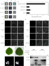
Fig. 5. Interactions between the CmLEC1 and CmC3H proteins.
a CmLEC1 and CmC3H; the negative control was pGADT7-T + pGBKT7, SD/-LW: SD/-Leu-Trp; SD/-LWHA: SD/-Leu-Trp-His-Ade. b The activity of β-galactosidase was determined by an enzyme assay. Data are the average value (±SD) of three independent experiments. Letters indicate a significant difference at P < 0.05, based on Student’s t test. c Interaction between CmLEC1 and CmC3H in the BiFC assays. Fluorescence was observed in the transformed cells, which resulted from complementation between the N-terminal region of GFP fused with CmLEC1 (CmLEC1-nGFP) and the C-terminal region of GFP fused with CmC3H (CmC3H-cGFP). The experiments were performed at least five times using different batches of N. benthamiana plants. d Interaction between CmLEC1 and CmC3H in the BiFC assays. Fluorescence was found in the transformed cells, which resulted from complementation between the C-terminal region of GFP fused with CmLEC1 (CmLEC1-cGFP) and the N-terminal region of GFP fused with CmC3H (CmC3H-nGFP). The experiments were performed at least five times using different batches of N. benthamiana plants. e Interaction between CmLEC1 and CmC3H in LCI assays. The LUC activity was determined 72 h later, using a CCD (charge coupled device) camera (Tanon 5200, China). f Interaction between CmLEC1 and CmC3H in an in vitro pull-down assay. The recombinant GST-CmLEC1 fusion was mixed with His-CmC3H fusion protein in equal volumes; following their incubation, the protein was purified by a GST column. In vitro-translated GST protein was used as a negative control. “Input” refers to the protein mixtures before the experiment; “Pull-down” means the purified protein mixture. The “+” indicates an existence, and the “−” indicates a non-existence. IB immunoblot
Fig. 6
Fig. 6. CmLEC1 and CmC3H synergistically activate the expression of CmLEA.
a Schematic diagram of upstream region corresponding to CmLEA folded-back structure. P1: 0 to −307 bp. P2: −307 to −607 bp. P3: −607 to −907 bp. P4: 0 to −907 bp. The lines below and above the blue box were fragments used in the yeast one-hybrid assays. Nucleotide substitutions in the wild-type cis element (CCAAT-box) and variant (P2m) of P2 are underlined. b The binding of CmLEC1 and CmLEA promoter in the yeast one-hybrid system was analyzed. The empty vector (pJG + pLacZi) was used as negative control. The CmLEA promoter was ligated to the pLacZi vector in yeast cells, and the ORF of CmLEC1 was cloned into pB42AD to obtain pB42AD-CmLEC1. c Schematic diagram of the double-reporter and effector plasmids for dual-luciferase (LUC) reporter assay. d, e CmLEC1 and CmC3H synergistically activated the expression of CmLEA in N. benthamiana leaves. In this experiment, a 0 to −907 bp promoter fragment of CmLEA was used; the constructs used in the assay are as shown above. Corresponding effectors and reporters were co-infiltrated into the tobacco leaves. d Representative images of fluorescence signals 3 days after infiltration are shown. e Relative LUC/REN ratios given were measured. The experiments were independently repeated three times and mean value ± SD are shown from three replicates
Fig. 7
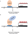
Fig. 7. Working model of the CmLEC1-mediated regulatory mechanism of embryo development in chrysanthemum.
CmLEC1 interacts with an embryo development factor, CmC3H, which together get bound to the upstream promoter region of CmLEA to positively promote the normal development of chrysanthemum embryos. (a) CmLEC1 gets bound to the upstream promoter region of CmLEA. (b) CmLEC1 interacts with an embryo development factor, CmC3H, which together get bound to the upstream promoter region of CmLEA

Similar articles

Cited by

References

    1. Kaul, M. L. H. Male Sterility in Higher Plants.Monographs on Theoretical and Applied Genetics (Springer, 1988).
    1. Zhou W, et al. A matter of life and death: molecular, physiological, and environmental regulation of seed longevity. Plant Cell Environ. 2019;43:293–302. doi: 10.1111/pce.13666. - DOI - PubMed
    1. Amlou M, Pla E, Moreteau B, David JR. Genetic analysis by interspecific crosses of the tolerance of drosophila sechelliato major aliphatic acids of its host plant. Genet. Sel. Evol. 1997;29:511–522. doi: 10.1186/1297-9686-29-4-511. - DOI - PubMed
    1. Rojas-Barrera IC, et al. Contemporary evolution of maize landraces and their wild relatives influenced by gene flow with modern maize varieties. Proc. Natl Acad. Sci. USA. 2019;116:21302–21311. doi: 10.1073/pnas.1817664116. - DOI - PMC - PubMed
    1. Burgess MB, et al. Effects of apomixis and polyploidy on diversification and geographic distribution in Amelanchier (Rosaceae) Am. J. Bot. 2014;101:1375–1387. doi: 10.3732/ajb.1400113. - DOI - PubMed