Review
. 2021 Aug 10;6(1):43.
doi: 10.1038/s41536-021-00153-z.

The bright side of fibroblasts: molecular signature and regenerative cues in major organs

Affiliations
Review

The bright side of fibroblasts: molecular signature and regenerative cues in major organs

Rita N Gomes et al. NPJ Regen Med. .

Erratum in

Abstract

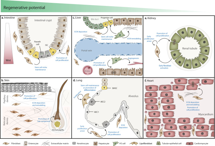
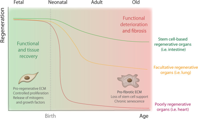
Fibrosis is a pathologic process characterized by the replacement of parenchymal tissue by large amounts of extracellular matrix, which may lead to organ dysfunction and even death. Fibroblasts are classically associated to fibrosis and tissue repair, and seldom to regeneration. However, accumulating evidence supports a pro-regenerative role of fibroblasts in different organs. While some organs rely on fibroblasts for maintaining stem cell niches, others depend on fibroblast activity, particularly on secreted molecules that promote cell adhesion, migration, and proliferation, to guide the regenerative process. Herein we provide an up-to-date overview of fibroblast-derived regenerative signaling across different organs and discuss how this capacity may become compromised with aging. We further introduce a new paradigm for regenerative therapies based on reverting adult fibroblasts to a fetal/neonatal-like phenotype.


Conflict of interest statement

The authors declare no competing interests.

Figures

Fig. 1
Fig. 1. The fibroblast as a mediator of regeneration in major organs.
a Intestinal fibroblasts are a component of the stem cell niche, secreting ECM, growth factors, and creating a Wnt gradient along the crypt promoting ISC proliferation/differentiation. b Dermal fibroblasts promote regeneration mainly by secreting specific ECM components, promoting wound healing. A specialized population of fibroblasts (dermal papilla) is also involved in hair neogenesis. c In the liver, portal fibroblasts and HS cells promote liver progenitor and hepatocyte proliferation after injury by multiple mechanisms. d Proliferative lung fibroblasts and lipofibroblasts promote regeneration by secreting structural ECM components and stimulating AEC2 cells with mitogens. e In the kidney, fibroblasts and epithelial cells communicate in a bidirectional fashion to coordinate tubular regeneration. f Specific ECM components putatively secreted by fibroblasts have been linked to cardiac regeneration in the neonatal heart.
Fig. 2
Fig. 2. Correlation of the regenerative potential of organs with aging.
Whilst less regenerative organs, as the heart, decrease their regenerative capacity abruptly after birth, regenerative organs, such as the skin and intestine, experience a progressive decline in their renewal capacity with increasing age. Organs like the lung depend on specific signals to mount a regenerative response—facultative regeneration—however, this ability is also impaired in the elderly. The loss of regenerative capacity in most organs seem to associate with the transition from a “young” to an “old” fibroblast phenotype. Whilst young fibroblasts are pro-regenerative and promote healthy tissue recovery, aged fibroblasts fail to support the regenerative niche, ultimately contributing to fibrosis and loss of organ function.

Similar articles

Cited by

References

    1. Wynn TA. Cellular and molecular mechanisms of fibrosis. J. Pathol. 2008;214:199–210. doi: 10.1002/path.2277. - DOI - PMC - PubMed
    1. Gurtner GC, Werner S, Barrandon Y, Longaker MT. Wound repair and regeneration. Nature. 2008;453:314–321. doi: 10.1038/nature07039. - DOI - PubMed
    1. Gonzalez AC, et al. Wound healing—a literature review. Bras. Dermatol. 2016;91:614–620. doi: 10.1590/abd1806-4841.20164741. - DOI - PMC - PubMed
    1. Henderson NC, Rieder F, Wynn TA. Fibrosis: from mechanisms to medicines. Nature. 2020;587:555–566. doi: 10.1038/s41586-020-2938-9. - DOI - PMC - PubMed
    1. Kendall RT, Feghali-Bostwick CA. Fibroblasts in fibrosis: novel roles and mediators. Front. Pharmacol. 2014;5:123. doi: 10.3389/fphar.2014.00123. - DOI - PMC - PubMed