. 2022 May;43(5):1324-1336.
doi: 10.1038/s41401-021-00749-7. Epub 2021 Aug 10.

Gout-associated monosodium urate crystal-induced necrosis is independent of NLRP3 activity but can be suppressed by combined inhibitors for multiple signaling pathways

Affiliations

Gout-associated monosodium urate crystal-induced necrosis is independent of NLRP3 activity but can be suppressed by combined inhibitors for multiple signaling pathways

Chun-Su Zhong et al. Acta Pharmacol Sin. 2022 May.

Abstract

Monosodium urate (MSU) crystals, the etiological agent of gout, are formed in joints and periarticular tissues due to long-lasting hyperuricemia. Although MSU crystal-triggered NLRP3 inflammasome activation and interleukin 1β (IL-1β) release are known to have key roles in gouty arthritis, recent studies revealed that MSU crystal-induced necrosis also plays a critical role in this process. However, it remains unknown what forms of necrosis have been induced and whether combined cell death inhibitors can block such necrosis. Here, we showed that MSU crystal-induced necrosis in murine macrophages was not dependent on NLRP3 inflammasome activation, as neither genetic deletion nor pharmacological blockade of the NLRP3 pathway inhibited the necrosis. Although many cell death pathways (such as ferroptosis and pyroptosis) inhibitors or reactive oxygen species inhibitors did not have any suppressive effects, necroptosis pathway inhibitors GSK'872 (RIPK3 inhibitor), and GW806742X (MLKL inhibitor) dose-dependently inhibited MSU crystal-induced necrosis. Moreover, a triple combination of GSK'872, GW806742X, and IDN-6556 (pan-caspase inhibitor) displayed enhanced inhibition of the necrosis, which was further fortified by the addition of MCC950 (NLRP3 inhibitor), suggesting that multiple cell death pathways might have been triggered by MSU crystals. Baicalin, a previously identified inhibitor of NLRP3, inhibited MSU crystal-induced inflammasome activation and suppressed the necrosis in macrophages. Besides, baicalin gavage significantly ameliorated MSU crystal-induced peritonitis in mice. Altogether, our data indicate that MSU crystals induce NLRP3-independent necrosis, which can be inhibited by combined inhibitors for multiple signaling pathways, highlighting a new avenue for the treatment of gouty arthritis.

Keywords: baicalin; inflammasome; monosodium urate crystals; necroptosis; regulated necrosis.


Conflict of interest statement

The authors declare no competing interests.

Figures
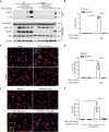
Fig. 1
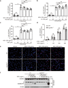
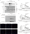
Fig. 1. MSU crystals induce NLRP3 inflammasome activation and IL-1β release.
Bone marrow-derived macrophages (BMDMs) from wild-type (WT) and NLRP3-deficient (NLRP3/−) mice were primed with lipopolysaccharide (LPS, 1 μg/mL) for 4 h, and then treated with indicated concentrations of monosodium urate (MSU) crystals for 6 h. a Western blot analysis was used to assess the expression levels of indicated proteins in cell lysates and culture supernatants. β-Actin was adopted as a loading control for cell lysates. Positive control (PC) was cell lysate from BMDMs primed with LPS (1 μg/mL) for 4 h followed by stimulation with adenosine triphosphate (ATP) (5 mM) for 30 min. b The levels of secreted IL-1β in culture supernatants were determined by cytometric bead array (CBA) assay. c, e Immunofluorescence microscopy was used to reveal ASC (red) distribution and nuclei (blue, stained by Hoechst 33342). LPS-primed WT BMDMs were pre-treated with or without MCC950 (1 μM, NLRP3 inhibitor) for 1 h, followed by MSU crystals (300 μg/mL) treatment for 6 h (e). Yellow arrows indicate ASC specks and enlarged insets show one cell with an ASC speck. Scale bars, 20 μm. d, f Percentages of cells with an ASC speck relative to total cells from 5 random fields. Data are shown as mean ± SD (n = 5). ***P < 0.001; CASP1, caspase-1; Pro-CASP1, pro-caspase-1
Fig. 2
Fig. 2. MSU crystal-induced necrosis does not require the activation of NLRP3 inflammasome.
BMDMs were treated as described in Fig. 1. LPS primed cells were pre-treated without or with indicated concentrations of MCC950 (a), VX-765 (b) (caspase-1 inhibitor), and disulfiram (DSF) (GSDMD inhibitor) (c) for 1 h, followed by co-stimulation with MSU crystals (300 μg/mL) for 6 h. Lytic cell death was assessed by propidium iodide (PI) uptake (red, indicating dead cells). PI-positive cells in five randomly chosen fields were quantified in (a–d). The percentage of lytic cell death is defined as the ratio of PI-positive cells relative to all cells. Data are shown as mean ± SD (n = 5). Representative PI and Hoechst 33342 (blue, indicating all cells) images from wild-type (WT) or NLRP3−/− cells captured by fluorescence microscopy are shown in (e). Scale bars, 20 μm. f GSDMD and GSDMD-NT levels in cell lysates were determined by Western blotting. β-Actin was used as a loading control. #denotes non-specific bands; ***P < 0.001, NS not significant
Fig. 3
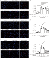
Fig. 3. MSU crystal-induced necrosis can be partly suppressed by inhibitors for the necroptosis pathway.
BMDMs from WT mice were first primed with LPS (1 μg/mL), then pre-treated without or with gradient doses of necrostatin-1 (Nec-1, RIPK1 inhibitor), GSK′872 (GSK, RIPK3 inhibitor), or GW806742X (GW, MLKL inhibitor) for 1 h, followed by stimulation with MSU crystals (300 μg/mL) for 6 h in the presence or absence of indicated inhibitors. Lytic cell death was measured by PI staining. a, c, and e Fluorescence microscopy was adopted to capture images with PI (red, staining dead cells) and Hoechst 33342 fluorescence (blue, staining all nuclei), Scale bars, 20 μm. b, d, and f The ratio of PI-positive cells was quantified. Data are shown as mean ± SD (n = 5). ***P < 0.001, NS not significant.
Fig. 4
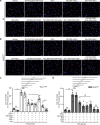
Fig. 4. Combination of multiple signaling pathway inhibitors can markedly suppress MSU crystal-induced necrosis in murine macrophages.
BMDMs from WT and NLRP3-/- mice were primed with LPS (1 μg/mL) for 4 h, and then pre-incubated without or with single or combination of multiple signaling pathway inhibitors for 1 h, followed by co-treatment with MSU crystals (300 μg/mL) for 6 h. a, b Representative lytic cell death images captured by fluorescence microscopy show PI (red, staining dead cells) combined with Hoechst 33342 (blue, staining all nuclei). Scale bars, 20 μm. c, d Quantitative percentage of PI-positive cells in a and b, respectively. Data are shown as mean ± SD (n = 5). *P < 0.05; **P < 0.01; ***P < 0.001, NS not significant. MCC950 (NLRP3 inhibitor, 1 μM), IDN-6556 (IDN, pan-caspase inhibitor, 20 μM), GSK′872 (GSK, RIPK3 inhibitor, 5 μM) and GW806742X (GW) (MLKL inhibitor, 1 μM) were used alone or in different combinations
Fig. 5
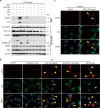
Fig. 5. p-MLKL is detected in macrophages treated with MSU crystals independent of NLRP3 inflammasome activation.
BMDMs from WT and NLRP3−/− mouse were primed with LPS (1 μg/mL) for 4 h and then pre-treated with one or a combination of multiple death pathway inhibitors for 1 h, followed by MSU (300 μg/mL) co-stimulation for 6 h. a Western blot analysis was used to detect the expression levels of indicated proteins in cell lysates and culture supernatants (Sup). β-Actin was used as a protein-loading control. b Immunofluorescence microscopy showing the distribution of p-MLKL (red) and ASC (green) while nuclei (blue) being revealed by Hoechst 33342 staining. IDN-6556 (IDN) (20 μM) alone was used to trigger necroptosis as a positive control. c Analysis of p-MLKL and ASC distribution in NLRP3/− BMDMs. White arrows and yellow arrowheads were respectively used to indicate ASC specks and p-MLKL fluorescence. Scale bars, 10 μm. #denotes non-specific bands.
Fig. 6
Fig. 6. Baicalin significantly inhibits not only NLRP3 inflammasome activation but also the necrosis in macrophages treated with MSU crystals.
BMDMs from WT mice were primed with LPS (1 μg/mL) for 4 h, and then pre-treated without or with graded doses of baicalin for 1 h, followed by stimulation with MSU crystals (300 μg/mL) for 6 h in the presence or absence of baicalin. a The expression levels of indicated proteins in cell lysates and culture supernatants (Sup) were analyzed by Western blotting. β-Actin was adopted as a loading control for cell lysates. b The levels of secreted IL-1β in culture supernatants were determined by cytometric bead array (CBA). c Western blotting analysis of ASC oligomers in Triton X-100 insoluble pellets cross-linked with disuccinimidyl suberate. d Lytic cell death was measured by lactic acid dehydrogenase (LDH) release in culture supernatants (n = 5). e Representative images showing PI (red, staining dead cells) combined with Hoechst 33342 (blue, staining all cells) fluorescence. Scale bars, 20 μm. f Quantification of PI-positive cells in 5 randomly chosen fields and ratio of PI-positive over all cells (revealed by Hoechst 33342) is defined as the percentages of lytic cell death. Data are shown as mean± SD (n = 5). *P < 0.05; **P < 0.01; ***P < 0.001, NS not significant.
Fig. 7
Fig. 7. MSU crystal-induced peritonitis can be ameliorated by baicalin gavage.
C57BL/6 J mice were administered intragastrically (i.g.) with baicalin (200 mg/kg body weight) or vehicle (2% Tween 80 in PBS) once a day for 2 consecutive days prior to intraperitoneal injection with sterile PBS (0.2 mL) or with 1 mg MSU crystals (in 0.2 mL sterile PBS). One hour later, mice were administered with vehicle or baicalin (i.g.) once again. Neutrophil and soluble cytokine levels in the peritoneal lavage fluids were quantified 4 h after MSU crystal administration (n = 5 mice per group). a Representative flow cytometric dot-plots of peritoneal exudate cells. b, c Quantification of neutrophils in peritoneal lavage fluids. d, e IL-1β and IL-6 levels in peritoneal lavage fluid supernatants were measured by cytometric bead array (CBA) assay. Data are shown as mean ± SD (n = 5). *P < 0.05, ***P < 0.001.

Similar articles

Cited by

References

    1. Dalbeth N, Merriman TR, Stamp LK. Gout. Lancet. 2016;388:2039–52. doi: 10.1016/S0140-6736(16)00346-9. - DOI - PubMed
    1. Desai J, Steiger S, Anders HJ. Molecular pathophysiology of gout. Trends Mol Med. 2017;23:756–68. doi: 10.1016/j.molmed.2017.06.005. - DOI - PubMed
    1. Dalbeth N, Choi HK, Joosten LAB, Khanna PP, Matsuo H, Perez-Ruiz F, et al. Gout. Nat Rev Dis Prim. 2019;5:69. doi: 10.1038/s41572-019-0115-y. - DOI - PubMed
    1. Mulay SR, Anders HJ. Crystallopathies. N Engl J Med. 2016;374:2465–76. doi: 10.1056/NEJMra1601611. - DOI - PubMed
    1. Martinon F, Pétrilli V, Mayor A, Tardivel A, Tschopp J. Gout-associated uric acid crystals activate the NALP3 inflammasome. Nature. 2006;440:237–41. doi: 10.1038/nature04516. - DOI - PubMed