. 2021 Aug 4:14:3471-3483.
doi: 10.2147/DMSO.S307771. eCollection 2021.

SERPINH1, Targeted by miR-29b, Modulated Proliferation and Migration of Human Retinal Endothelial Cells Under High Glucose Conditions

Affiliations

SERPINH1, Targeted by miR-29b, Modulated Proliferation and Migration of Human Retinal Endothelial Cells Under High Glucose Conditions

Lingfei Hu et al. Diabetes Metab Syndr Obes. .

Abstract

Aim: In the present study, we performed bioinformatics studies and in vitro functional assays to explore the underlying role of serpin family H member 1 (SERPINH1) in the diabetic retinopathy.

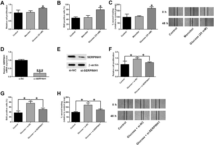
Methods: Common differentially expressed genes (DEGs) between diabetic retinal tissues and normal retinal tissues were analyzed using Gene Expression Omnibus (GEO) database. The proliferation and migration of human retinal endothelial cells (HRECs) was evaluated by MTS, EdU and wound healing assays, respectively; the miRNA and mRNAs expression levels of hub genes in HRECs were determined using quantitative real-time PCR (qRT-PCR). Protein levels were determined using a Western blot assay.

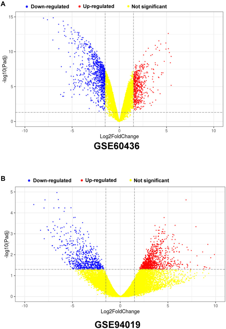
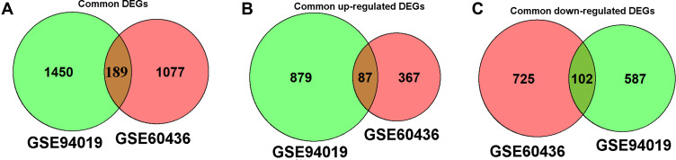
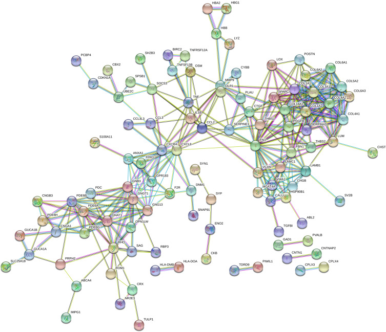
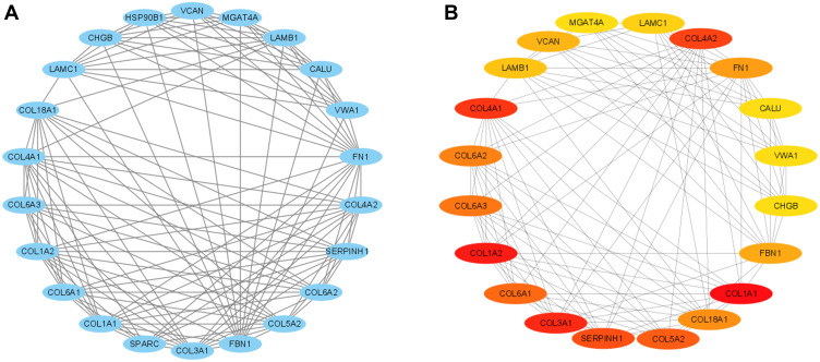
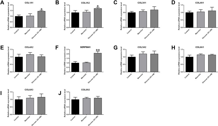
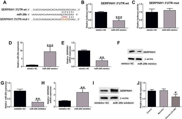
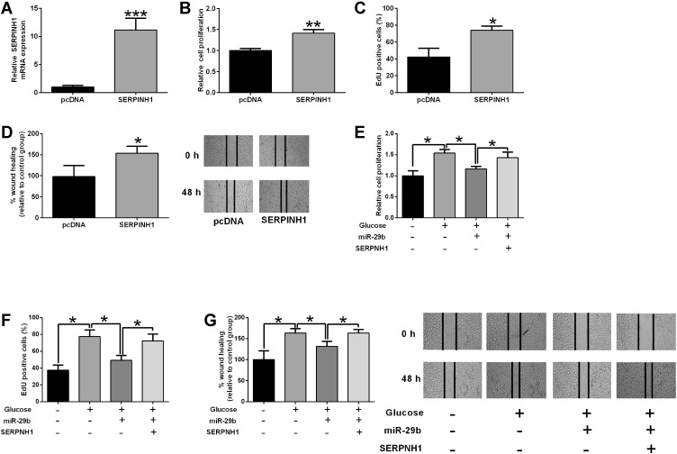
Results: A total of 189 common DEGs were screened between two GEO datasets (GSE60436 and GSE94019), and ten potential hub genes that may link to the progression of diabetic retinopathy were detected. The qRT-PCR results showed that collagen, type I, alpha 1 (COL1A1), Collagen, type I, alpha 2 (COL1A2) and serpin family H member 1 (SERPINH1) mRNA expression levels were up-regulated in the HRECs after being exposed to high glucose for 48 h. Silence of SERPINH1 repressed the high glucose-induced increase in proliferation and migration of HRECs. SERPINH1 was a target of miR-29b and was suppressed by miR-29 in HRECs. SERPINH1 overexpression promoted HREC proliferation and migration. Furthermore, miR-29b suppressed HREC proliferation and migration under high-glucose stimulation, which was significantly attenuated by enforced expression of SERPINH1.

Conclusion: In conclusion, by performing the integrated bioinformatics analysis, the present study suggested that 3 hub genes (COL1A1, COL1A2 and SERPINH1) may be associated with diabetic retinopathy pathophysiology. Further mechanistic studies indicated that miR-29b/SERPINH1 signaling participated in high glucose-induced enhancement in the proliferation and migration of HRECs.

Keywords: HRECs; SERPINH1; bioinformatics analysis; diabetic retinopathy; migration; proliferation.


Conflict of interest statement

The authors report no conflicts of interest in this work.

Figures

Figure 1
Figure 1
Volcano plots of the DEGs in the GSE60436 and GSE94019 datasets. (A) Volcano plot of DEGs from GSE60436; (B) Volcano plot of DEGs from GSE94019.
Figure 2
Figure 2
Venn diagram showing the common DEGs between GSE60436 and GSE94019 datasets. (A) Venn diagram of the common DEGs between GSE60436 and GSE94019. (B) Venn diagram of the common up-regulated DEGs between GSE60436 and GSE94019. (C) Venn diagram of the common down-regulated DEGs between GSE60436 and GSE941919.
Figure 3
Figure 3
PPI network of the DEGs.
Figure 4
Figure 4
Identification of hub gene modules using Cytoscape. (A) Module of PPI network constructed by MCODE. (B) Module of PPI network constructed by CytoHubba.
Figure 5
Figure 5
Effects of high glucose treatment on the hub genes in the HRECs. The mRNA expression levels of (A) COL1A1, (B) COL1A2, (C) COL3A1, (D) COL4A1, (E) COL4A2, (F) SERPINH1, (G) COL5A2, (H) COL6A1, (I) COL6A3 and (J) COL6A2 in the HRECs after different treatments were determined by qRT-PCR. *p<0.05 and **p<0.01.
Figure 6
Figure 6
SERPINH1 silence attenuated the high glucose-induced increase in cell proliferation and migration of HRECs. (A) The cell proliferation of HRECs after different treatments (control, mannitol, 25 mM glucose) was determined by CCK-8 assay. (B) EdU assay was used to determined HREC proliferation after different treatments (control, mannitol, 25 mM glucose). (C) The HREC migration after different treatments (control, mannitol, 25 mM glucose) was determined by wound healing assay. (D) The mRNA and (E) protein expression levels of SERPINH1 in HRECs after being transfected with si-NC or si-SERPINH1 were determined by qRT-PCR and Western blot assay, respectively. (FH) HRECs were transfected with si-NC or si-SERPINH1 followed by treating with 25 mM glucose, the cell proliferation and migration of HRECs were determined by CCK-8, EdU and wound healing assay, respectively. *p<0.05.
Figure 7
Figure 7
MiR-29b repressed the expression of SERPINH1 in HRECs. (A) The predicted complementary sequences between miR-29b and SERPINH1 3ʹUTR. (B and C) The luciferase reporter activities of SERPINH1 3ʹUTR-wt (B) and SERPINH1 3ʹUTR-mut (C) in HRECs after being transfected with mimics NC or miR-29b mimics were determined by Dual-Luciferase Reporter assay. (D) The miR-29b expression level in HRECs after being transfected with mimics NC or miR-29b mimics were determined by qRT-PCR. (E and F) The mRNA and protein expression levels of SERPINH1 in HRECs after being transfected with mimics NC or miR-29b mimics were determined by qRT-PCR. (G) The miR-29b expression level in HRECs after being transfected with inhibitor NC or miR-29b inhibitor was determined by qRT-PCR. (H and I) The mRNA and protein expression levels of SERPINH1 in HRECs after being transfected with inhibitor NC or miR-29b inhibitor were determined by qRT-PCR. (J) The mRNA expression levels of miR-29b in the HRECs after different treatments were determined by qRT-PCR. *p<0.05, **p<0.01 and ***p<0.001.
Figure 8
Figure 8
MiR-29b/SERPINH1 axis participated in the glucose-induced increase in HREC proliferation and migration. (A) The mRNA expression level of SERPINH1 in HRECs after being transfected with pcDNA3.1 or pcDNA3.1-SERPINH1 was determined by qRT-PCR. (BD) HRECs were transfected with pcDNA3.1 or pcDNA3.1-SERPINH1, the cell viability, proliferation and migration of HRECs were determined by MTS (B), EdU (C) and wound healing assay (D), respectively. (EG) HRECs were transfected with different miRNAs or plasmids followed by treatment with 25 mM glucose, the cell viability, proliferation and migration of HRECs were determined by MTS, EdU and wound healing assay, respectively. *p<0.05, **p<0.01 and ***p<0.001.

Similar articles

Cited by

References

    1. Amoaku WM, Ghanchi F, Bailey C, et al. Diabetic retinopathy and diabetic macular oedema pathways and management: UK Consensus Working Group. Eye. 2020;34(Suppl 1):1–51. doi:10.1038/s41433-020-0961-6 - DOI - PMC - PubMed
    1. Liu Y, Li J, Ma J, Tong N. The threshold of the severity of diabetic retinopathy below which intensive glycemic control is beneficial in diabetic patients: estimation using data from large randomized clinical trials. J Diabetes Res. 2020;2020:8765139. doi:10.1155/2020/8765139 - DOI - PMC - PubMed
    1. Ahmad I, Hoda M. Attenuation of diabetic retinopathy and neuropathy by resveratrol: review on its molecular mechanisms of action. Life Sci. 2020;245:117350. doi:10.1016/j.lfs.2020.117350 - DOI - PubMed
    1. Singh RR. Next-generation sequencing in high-sensitive detection of mutations in tumors: challenges, advances, and applications. J Mol Diagn. 2020;22(8):994–1007. doi:10.1016/j.jmoldx.2020.04.213 - DOI - PubMed
    1. Lam JD, Oh DJ, Wong LL, et al. Identification of RUNX1 as a mediator of aberrant retinal angiogenesis. Diabetes. 2017;66(7):1950–1956. doi:10.2337/db16-1035 - DOI - PMC - PubMed