. 2021 Aug;10(10):e12129.
doi: 10.1002/jev2.12129. Epub 2021 Aug 2.

Cryptococcus extracellular vesicles properties and their use as vaccine platforms

Affiliations

Cryptococcus extracellular vesicles properties and their use as vaccine platforms

Juliana Rizzo et al. J Extracell Vesicles. 2021 Aug.

Abstract

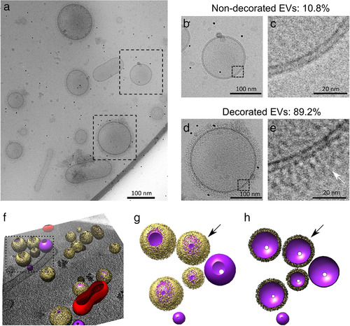
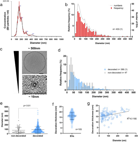
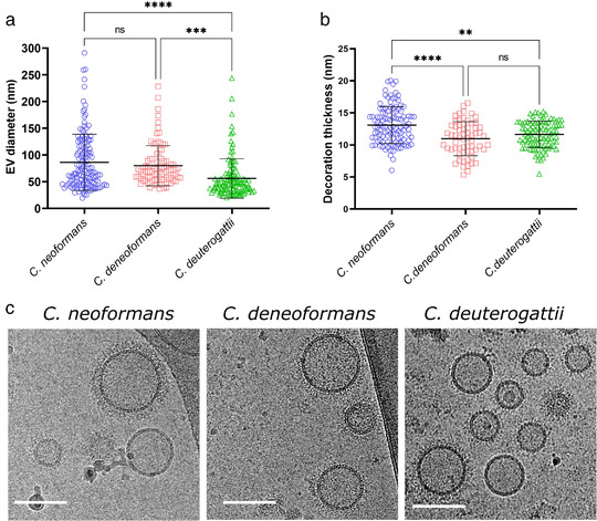
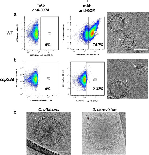
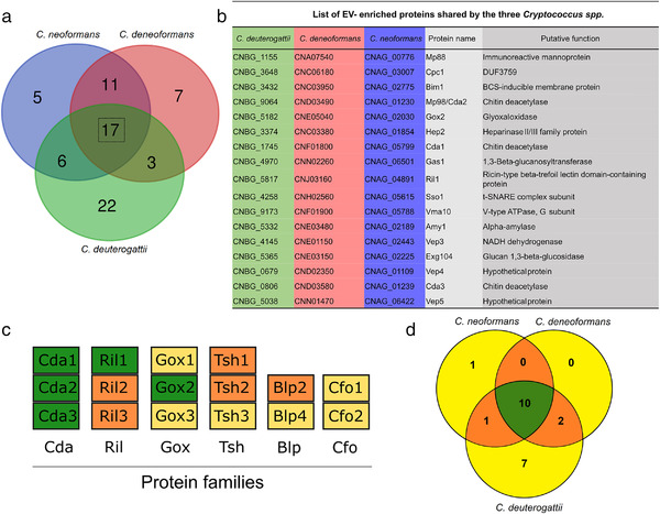
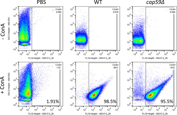
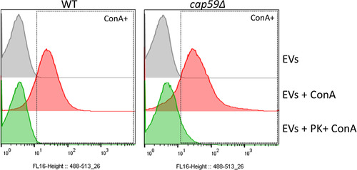
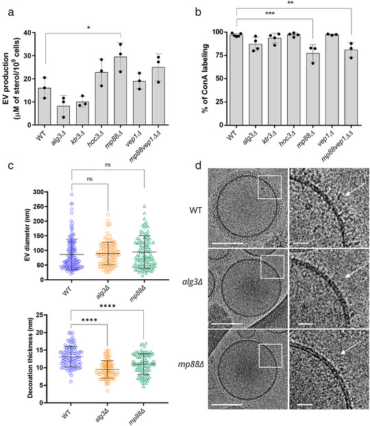
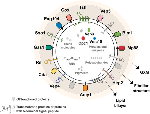
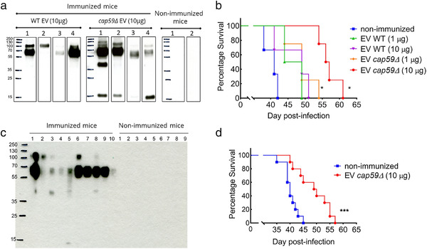
Whereas extracellular vesicle (EV) research has become commonplace in different biomedical fields, this field of research is still in its infancy in mycology. Here we provide a robust set of data regarding the structural and compositional aspects of EVs isolated from the fungal pathogenic species Cryptococcus neoformans, C. deneoformans and C. deuterogattii. Using cutting-edge methodological approaches including cryogenic electron microscopy and cryogenic electron tomography, proteomics, and flow cytometry, we revisited cryptococcal EV features and suggest a new EV structural model, in which the vesicular lipid bilayer is covered by mannoprotein-based fibrillar decoration, bearing the capsule polysaccharide as its outer layer. About 10% of the EV population is devoid of fibrillar decoration, adding another aspect to EV diversity. By analysing EV protein cargo from the three species, we characterized the typical Cryptococcus EV proteome. It contains several membrane-bound protein families, including some Tsh proteins bearing a SUR7/PalI motif. The presence of known protective antigens on the surface of Cryptococcus EVs, resembling the morphology of encapsulated virus structures, suggested their potential as a vaccine. Indeed, mice immunized with EVs obtained from an acapsular C. neoformans mutant strain rendered a strong antibody response in mice and significantly prolonged their survival upon C. neoformans infection.

Keywords: Cryo‐EM; cryptococcus; extracellular vesicles; fungal infections; mannoproteins; vaccine.


Figures

FIGURE 1
FIGURE 1
Cryo‐electron microscopy analysis of C. neoformans extracellular vesicles (EVs). Cryo‐EM analysis revealed a heterogeneous population of vesicles with diverse structural aspects, previously unappreciated in fungal EVs (a). As shown, the EVs were delimitated by a lipid bilayer (b to e), which showed either no decoration (in 10.8% vesicles, panels b and c) or a fibrillar decoration (arrows) in 89.2% of the EVs analysed (panels d and e). Three‐dimensional organization of the fibrillar decoration (yellow) on the membrane (purple) of EVs as revealed by cryo‐electron tomography analysis (f), magnified in panels g and h. Full surface representation models as seen from top view (g). Same models clipped with clipping plane oriented perpendicular to line of sight (h). Data presented in this figure have been generated using images obtained using a Titan Krios (Thermo Scientific) transmission electron microscope
FIGURE 2
FIGURE 2
Analysis of size and structural diversity of C. neoformans EVs. NTA analysis of purified EVs revealed a size diameter ranging from 80 to 500 nm, with the highest distribution around 150 nm (a). Frequency distribution of EV diameters determined by CryoEM, a total of 434 regular EVs were analysed. The analysis based on CryoEM tomograms revealed a wider range of EV size distribution, from 10 to 500 nm diameter, with the highest relative frequency below 100 nm (b). Cryo‐EM images exemplifying EV size range. Scale bars corresponding to 100 nm (c). EV size distribution according to the presence or absence of surface decoration (d). Non‐decorated EVs have a smaller diameter size distribution compared to decorated ones (e). Analysis of decoration thickness from Cryo‐EM images from 105 single EVs (f). Analysis of a potential relationship between decoration thickness and EV diameter by linear regression (g). Data presented in this figure have been generated using images obtained using a Titan Krios (Thermo Scientific) transmission electron microscope. Error bars show means ± SD. Sample size (n) is indicated and, in brackets, the number of vesicles in that category that exceeded 500 nm in size
FIGURE 3
FIGURE 3
Comparative analysis of size and structural diversity of EVs in C. neoformans, C. deneoformans and C. deuterogattii. Analysis of EV diameters revealed a smaller size distribution in C. deuterogattii strain R265 than in C. neoformans KN99α and C. deneoformans strain JEC21. The total numbers of vesicles analysed were C. neoformans (n = 143 for size and n = 112 for decoration), C. deneoformans (n = 90 for size and n = 63 for decoration), C. deuterogattii (n = 115 for size and n = 95 for decoration) (a). Analysis of the decoration thickness revealed a smaller distribution for C. deneoformans and C. deuterogattii compared with C. neoformans (b). Illustrative images of size and decoration of EVs obtained from the three species. The data presented in this figure have been generated using images obtained using a TECNAI F20 transmission electron microscope (c). Error bars show means ± SD. Scale bars represent 100 nm
FIGURE 4
FIGURE 4
Flow cytometry analysis of C. neoformans EVs incubated with monoclonal anti‐GXM antibody. FACS analysis of wild type (WT) and the acapsular cap59Δ EVs in PBS (‐ mAb anti‐GXM) or in the presence of the monoclonal antibody raised against the capsular polysaccharide 18b7 (+ mAb anti‐GXM) (a). The analysis revealed strong labelling of WT vesicles (74.7%), compared to the weak labelling in the mutant (2.33%), (b). Despite the important labelling difference, C. neoformans WT and cap59Δ strains released EVs bearing similar surface decoration, shown by the cryo‐EM (arrows), as well as EVs obtained from other fungal species such as C. albicans and S. cerevisiae (c). These cryo‐EM data have been generated using a TECNAI F20 transmission electron microscope. Scale bar represents 100 nm. This experiment was repeated twice with similar results
FIGURE 5
FIGURE 5
Analysis of Cryptococcus spp protein cargo. Venn diagram revealing shared and unique EV‐associated proteins in C. neoformans, C. deneoformans, and C. deuterogattii. Seventeen proteins were identified to be associated with EVs in all three Cryptococcus species (a). List of the gene loci and the corresponding proteins commonly found in EVs released by the three species, which could be considered as putative cryptococcal EV‐protein markers (b). Most of the proteins are predicted to be either GPI‐anchored proteins, to contain a signal peptide or to possess other membrane domains, according to preGPI, signalP and TMHMM website, respectively. Six protein families appeared to be typical of Cryptococcus EVs, including the Chitin deacetylase family (Cda), the Ricin‐type beta‐trefoil lectin domain‐containing protein family (Ril), the putative glyoxal oxidase family (Gox), the tetraspanin membrane proteins containing a SUR7/PalI family motif (Tsh), the pr4/barwin domain protein family (Blp), and the multicopper oxidase (Cfo). Among these families, the proteins present in all three species are shown in green, proteins present in two species in orange and proteins present in only one species in yellow (c). We also identify 21 putative GPI‐anchored proteins, as predicted by PredGPI, and 10 of them were present in all three species (d)
FIGURE 6
FIGURE 6
Flow cytometry analysis of C. neoformans EVs incubated with GFP‐labelled ConA. FACS analysis of EVs obtained from C. neoformans wild type and cap59Δ cells. EVs were incubated with GFP‐labelled ConA lectin. After ultracentrifuge washing, the EV pellets were mixed in BD Trucount tubes (BD Biosciences), containing a known number of fluorescent beads as internal control. The number of events for each reading was fixed to 100,000 events and the percentage and intensity of ConA labelling were recorded. This experiment was repeated three times with similar results
FIGURE 7
FIGURE 7
EV proteinase K treatment reduces ConA binding. FACS analysis of EVs obtained from C. neoformans WT and cap59Δ cells after proteinase K treatment. Proteinase K‐treated EVs were submitted to ConA labelling, ultracentrifuge washed and analysed by flow cytometry. EV pellets were mixed in BD Trucount tubes (BD Biosciences), containing a known number of fluorescent beads as an internal control. The number of events for each reading was fixed to 100,000 events and the percentage and intensity of ConA labelling were recorded. EVs treated using the same protocol but omitting the enzyme were used as controls
FIGURE 8
FIGURE 8
Analysis of C. neoformans mutant strain EVs. Evaluation of EV production by the different mutant strains as estimated by the measure of the sterol concentration using the Amplex™ Red Cholesterol Assay Kit (a). Impact of the different mutations on the percentage of ConA‐labelled EVs as estimated through flow cytometry (b). Analysis of EV size diameter in the mp88Δ and alg3Δ mutant strains as compared to the wild type (WT). The total number of vesicles analysed were WT (n = 143 for size and n = 112 for decoration), mp88Δ (n = 107 for size and n = 86 for decoration), alg3Δ (n = 119 for size and n = 92 for decoration) (c). Analysis of the decoration thickness revealed a smaller distribution associated with ALG3 or MP88 deletions, as exemplified by illustrative images from the three strains (d). The cryo‐EM images were obtained using a TECNAI F20 transmission electron microscope. ConA labelling and sterol measurements were done for at least three independent biological replicates Error bars are represented as means ± SD. Scale bars represent 100 nm and 20 nm in magnified views of boxed areas in D
FIGURE 9
FIGURE 9
Model of simplified molecular structure and composition of Cryptococcus EVs. In accordance with previous reports and in the light of our data, a new model of Cryptococcus EVs is suggested, where the outer layer is composed of the capsular polysaccharide glucuronoxylomannan (GXM), and the lipid bilayer is covered by many proteins, including mannoproteins, making the visible fibrillar structure resolved by cryo‐EM. Most of the proteins are predicted to be GPI‐anchored, to contain a signal peptide or to possess other membrane domains, according to preGPI, signalP and TMHMM, respectively. Three proteins, the hypothetical protein Cpc1, the putative V‐type ATPase (Vma10) and the Vep3 are predicted to be soluble. It is still unclear if these proteins are indeed inside the vesicular lumen or linked to another transmembrane protein. For simplification, the lipid content was not explored, but previous works shown the presence of sterol, phospholipoids and sphingolipids. Additionally, Cryptococcus EVs were also described to contain other cargoes, such as RNA, pigments, small molecules, and polysaccharides, including GXM, as detailed in plain text
FIGURE 10
FIGURE 10
Vaccination assays using C. neoformans EVs. Female 6‐weeks old BALB/c mice were immunized with C. neoformans EVs via intraperitoneal injection, followed by intranasal infection with 1 × 104 yeasts of wild type (WT) C. neoformans, and the mouse survival was monitored. In the first pilot experiment, mice (n = 4 per group) were immunized with EVs from wild type or cap59Δ strain (1 and 10 μg in 100 μl of PBS) and control mice were injected with 100 μl PBS. Western Blot using mouse sera against fungal EV confirmed that all immunized mice produced antibodies against EV proteins (a). All EV‐immunized mice survived longer than the non‐immunized ones, but the immunization with cap59Δ EVs rendered a significantly prolonged mouse survival (*P = 0.01) (b). For the second set of experiment, mice (n = 10 per group) were immunized with EVs from cap59Δ mutant strain (10 μg/100 μl in PBS) and control mice were injected with 100 μl PBS. Again, Western blot using mouse sera against fungal EVs confirmed that all immunized mice produced antibodies against EV proteins (c). EV‐immunized mice showed significantly prolonged survival (*P = 0.0006) compared to the non‐immunized group (d). Comparison of the survival curves was made by GraphPad Prism 9, using the Log‐rank (Mantel‐Cox) test

References

    1. Albuquerque, P. C. , Nakayasu, E S. , Rodrigues, M L. , Frases, S. , Casadevall, A. , Zancope‐Oliveira, R. M. , Almeida, I. C. , & Nosanchuk, J. D. (2008). Vesicular transport in Histoplasma capsulatum: An effective mechanism for trans‐cell wall transfer of proteins and lipids in ascomycetes. Cellular Microbiology, 10, 1695–1710. - PMC - PubMed
    1. Armenteros, A. , Juan, J. , Tsirigos, K. D. , Sønderby, C. K. , Petersen, T. N. , Winther, O. , Brunak, S. , von Heijne, G. , & Nielsen, H. (2019). SignalP 5.0 improves signal peptide predictions using deep neural networks. Nature Biotechnology, 37, 420–423. - PubMed
    1. Arraud, N. , Linares, R. , Tan, S. , Gounou, C. , Pasquet, J. M. , Mornet, S. , & Brisson, A. R. (2014). Extracellular vesicles from blood plasma: Determination of their morphology, size, phenotype and concentration. Journal of Thrombosis and Haemostasis, 12, 614–627. - PubMed
    1. Bachurski, D. , Schuldner, M. , Nguyen, P. ‐. H. , Malz, A. , Reiners, K. S. , Grenzi, P. C. , Babatz, F. , Schauss, A. C. , Hansen, H. P. , Hallek, M. , & Strandmann, E. P. (2019). Extracellular vesicle measurements with nanoparticle tracking analysis ‐ An accuracy and repeatability comparison between NanoSight NS300 and ZetaView. Journal of Extracellular Vesicles, 8, 1596016–16. - PMC - PubMed
    1. Baker, L G. , Specht, C A. , & Lodge, J K. (2011). Cell wall chitosan is necessary for virulence in the opportunistic pathogen cryptococcus neoformans , Eukaryotic Cell, 10, 1264. - PMC - PubMed

Publication types

MeSH terms