The survival of non-traumatic osteonecrosis of femoral head at ARCO II with ring-shaped sclerotic zone: a mid-term follow-up retrospective study
- PMID: 34377513
- PMCID: PMC8349575
- DOI: 10.1093/jhps/hnab013
The survival of non-traumatic osteonecrosis of femoral head at ARCO II with ring-shaped sclerotic zone: a mid-term follow-up retrospective study
Abstract
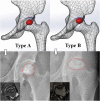
The sclerotic zone in the osteonecrosis of femoral head (ONFH), containing condensed trabecular bone and abundant neovascularization, is the transition area between osteonecrosis and normal tissue. Due to the prominent feature in ONFH, the characteristics of the sclerotic zone might indicate the femoral head survival of the disease. Thirty ONFH patients (41 hips) with ring-shaped sclerotic zone at Association Research Circulation Osseous-II were recruited during 1996 to 2019, and the corresponding radiographic images in their follow-up are reviewed retrospectively. Two subtypes (type A and B) are defined to discriminate different locations of ring-shaped sclerotic zone in the femoral head (center or subchondral bone plate) in accordance with the radiographic images. The natural history of the enrolled subjects was followed up for average 9 years to record and compare their collapse incidences as well as the progress of hip symptoms. Chi-square test shows that the occurrence rates of symptomatic hip of type A are significantly lower than that of type B and differences between these two groups were significant (P < 0.05). Kaplan Meier survival curve analysis shows that the mean survival time of type A is 247.600 M (95% CI: 203.072 ∼ 292.128 M) and type B is 88.795 M (95% CI: 72.607 ∼ 104.984 M). The survival rate of femoral head of type A is significantly higher than that of type B (P < 0.005). This study demonstrates that type A shows a more satisfactory clinical outcomes and lower femoral head collapse rate in a mid-term follow-up.
© The Author(s) 2021. Published by Oxford University Press.
Figures


Similar articles
-
[EFFECTIVENESS OF BONE GRAFTING THROUGH WINDOWING AT FEMORAL HEAD-NECK JUNCTION FOR TREATMENT OF OSTEONECROSIS WITH SEGMENTAL COLLAPSE OF FEMORAL HEAD].Zhongguo Xiu Fu Chong Jian Wai Ke Za Zhi. 2016 Apr;30(4):397-401. Zhongguo Xiu Fu Chong Jian Wai Ke Za Zhi. 2016. PMID: 27411263 Chinese.
-
[Clinical significance of different imaging manifestations of osteonecrosis of femoral head in the peri-collapse stage].Zhongguo Xiu Fu Chong Jian Wai Ke Za Zhi. 2021 Sep 15;35(9):1105-1110. doi: 10.7507/1002-1892.202103221. Zhongguo Xiu Fu Chong Jian Wai Ke Za Zhi. 2021. PMID: 34523274 Free PMC article. Chinese.
-
The Therapeutic Effect of Huo Xue Tong Luo Capsules in Association Research Circulation Osseous (ARCO) Stage II Osteonecrosis of the Femoral Head: A Clinical Study With an Average Follow-up Period of 7.95 Years.Front Pharmacol. 2021 Nov 23;12:773758. doi: 10.3389/fphar.2021.773758. eCollection 2021. Front Pharmacol. 2021. PMID: 34899331 Free PMC article.
-
Hip Arthroscopy Debridement Combined with Multiple Small-Diameter Fan-Shaped Low-Speed Drilling Decompression in the Treatment of Early and Middle Stage Osteonecrosis of the Femoral Head: 14 Years Follow-Up.Orthop Surg. 2024 Mar;16(3):604-612. doi: 10.1111/os.13985. Epub 2024 Jan 23. Orthop Surg. 2024. PMID: 38263763 Free PMC article.
-
Mid- to long-term results of modified avascular fibular grafting for ONFH.J Hip Preserv Surg. 2021 Oct 18;8(3):274-281. doi: 10.1093/jhps/hnab046. eCollection 2021 Aug. J Hip Preserv Surg. 2021. PMID: 35414946 Free PMC article. Review.
Cited by
-
Lateral classification system predicts the collapse of JIC type C1 nontraumatic osteonecrosis of the femoral head: a retrospective study.BMC Musculoskelet Disord. 2023 Sep 26;24(1):757. doi: 10.1186/s12891-023-06890-0. BMC Musculoskelet Disord. 2023. PMID: 37749534 Free PMC article.
-
Sclerotic zone in femoral head necrosis: from pathophysiology to therapeutic implications.EFORT Open Rev. 2023 Jun 8;8(6):451-458. doi: 10.1530/EOR-22-0092. EFORT Open Rev. 2023. PMID: 37289132 Free PMC article. Review.
References
-
- Fukushima W, Yamamoto T, Takahashi S et al. The effect of alcohol intake and the use of oral corticosteroids on the risk of idiopathic osteonecrosis of the femoral head: a case-control study in Japan. Bone Joint J 2013; 95-B: 320e5–325. - PubMed
-
- Yoon B-H, Jones LC, Chen C-H et al. Etiologic classification criteria of ARCO on femoral head osteonecrosis part 1: glucocorticoid-associated osteonecrosis. J Arthroplast 2019; 34: 163e168–e1. - PubMed
LinkOut - more resources
Full Text Sources

