Review
. 2021 Jul 8;6(7):593-606.
doi: 10.1302/2058-5241.6.200042. eCollection 2021 Jul.

Ankle fractures in children

Affiliations
Review

Ankle fractures in children

Benjamín Cancino et al. EFORT Open Rev. .

Abstract

Ankle fractures are common in children, and they have specific implications in that patient population due to frequent involvement of the physis in a bone that has growth potential and unique biomechanical properties.Characteristic patterns are typically evident in relation to the state of osseous development of the segment, and to an extent these are age-dependent.In a specific type known as transitional fractures - which occur in children who are progressing to a mature skeleton -a partial physeal closure is evident, which produces multiplanar fracture patterns.Computed tomography should be routine in injuries with joint involvement, both to assess the level of displacement and to facilitate informed surgical planning.The therapeutic objectives should be to achieve an adequate functional axis of the ankle without articular gaps, and to protect the physis in order to avoid growth alterations.Conservative management can be utilized for non-displaced fractures in conjunction with strict radiological monitoring, but surgery should be considered for fractures involving substantial physeal or joint displacement, in order to achieve the therapeutic goals. Cite this article: EFORT Open Rev 2021;6:593-606. DOI: 10.1302/2058-5241.6.200042.

Keywords: Salter–Harris fractures of the tibia; paediatric ankle; physeal ankle fracture.


Conflict of interest statement

ICMJE Conflict of interest statement: The author declares no conflict of interest relevant to this work.

Figures

Fig. 1
Fig. 1
X-rays of both legs of an eight-month-old boy. Bilateral metaphyseal impaction is evident. Investigation in accordance with a standard protocol suggested that the injuries were non-accidental.
Fig. 2
Fig. 2
X-rays of a newborn’s left ankle. Physis is evident transversely in the tibia and fibula, the latter being more distal, at the level of the tibial epiphysis.
Fig. 3
Fig. 3
X-rays of a 12-month-old boy’s right ankle. Secondary ossification nuclei are present in the distal epiphyses of the tibia and fibula.
Fig. 4
Fig. 4
X-rays of a three-year-old girl’s right ankle. Anteromedial undulation of the tibial physis is evident, and the fibular physis reaches the tibial articular surface.
Fig. 5
Fig. 5
X-rays of a seven-year-old girl’s left ankle. The beginning of ossification of the medial malleolus is evident, as is distal ossification of the lateral malleolus.
Fig. 6
Fig. 6
X-rays of the right ankle of an 11-year-old boy. The beginning of physeal closure is visible at the level of Poland’s hump.
Fig. 7
Fig. 7
Sequence of distal physeal closure of the tibia. Closure begins from the anteromedial area (A), then progresses medially (B), posteriorly (C), and laterally (D).
Fig. 8
Fig. 8
X-rays of the left ankle of a 10-year-old girl with a metaphyseal bending fracture (arrows).
Fig. 9
Fig. 9
X-rays of the right ankle of a 10-year-old girl. Park–Harris lines (arrows) that are parallel to the physis are visible, indicating normal growth.
Fig. 10
Fig. 10
Radiography (A) and computed tomography (B, C) of the right ankle of an 11-year-old girl with a Salter–Harris type III fracture with joint involvement.
Fig. 11
Fig. 11
Johnson and Fahl classification: 1, abduction type; 2, plantar flexion type; 3, adduction type.
Fig. 12
Fig. 12
Salter–Harris classification: type I, complete separation of the epiphysis from the metaphysis through the physis; type II, fracture line along the physis and a portion of metaphysis; type III, fracture line along the physis and a portion of epiphysis; type IV, fracture line through epiphysis, physis and metaphysis; type V, crushing force through the epiphysis to the physis.
Fig. 13
Fig. 13
Dias-Tachdjian classification: (A) supination-inversion; (B) supination-plantar flexion; (C) supination-external rotation; (D) pronation-eversion; (E) Salter–Harris III of distal tibial epiphysis; (F) triplane fracture.
Fig. 14
Fig. 14
Computed tomography of the left ankle of a 12-year-old boy with a classic pattern triplane fracture. Epiphyseal fracture is visible in the coronal section and in the distal axial section (A, B). Metaphyseal fracture is visible in the proximal axial section and in the sagittal section (C, D). Physeal compromise is evident in the coronal and sagittal sections (A, D).
Fig. 15
Fig. 15
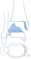
Triplanar fracture patterns. Lateral epiphyseal in two parts (A), lateral epiphyseal in three parts (B), lateral epiphyseal in four parts (C), and medial epiphyseal in three parts (D).
Fig. 16
Fig. 16
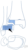
Extra-articular triplanar fracture pattern.
Fig. 17
Fig. 17
Computed tomography of the left ankle of a 13-year-old girl with an extra-articular triplanar fracture. A sagittal fracture that compromises the medial malleolus is visible in the coronal (A) and axial sections (B), and a coronal metaphyseal pattern is evident in the sagittal section (C).
Fig. 18
Fig. 18
Juvenile Tillaux fracture pattern. The anterior tibiofibular ligament is inserted into the avulsed fragment of the anterior distal tubercle of the tibia (arrow).
Fig. 19
Fig. 19
Computed tomography of the right ankle of a 15-year-old boy depicting a juvenile Tillaux fracture. A square-shaped fragment is observed in all sections.
Fig. 20
Fig. 20
Radiography (A) and computed tomography (B, C) of the right ankle of an 11-year-old boy with a Salter–Harris type II fracture, depicting a posterolateral Thurston Holland fragment (dotted blue line on the radiograph).
Fig. 21
Fig. 21
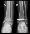
Right ankle of a 13-year-old boy with a displaced Salter–Harris type II fracture (A), and subsequent postoperative control after fixation with two laterally crossed smooth K-wires (B).
Fig. 22
Fig. 22
Left ankle of a 12-year-old boy with a medial malleolus Salter–Harris type IV fracture (A), and postoperative control after performing open reduction internal fixation with a cannulated screw, without compromising the physis (B).
Fig. 23
Fig. 23
Right ankle of a 14-year-old girl with a juvenile Tillaux fracture with anterior displacement (A, B). Surgical management was performed using open reduction internal fixation with a transphyseal cannulated screw (C).
Fig. 24
Fig. 24
Anteroposterior view of the right ankle of a 13-year-old boy with a triplanar fracture with a Gothic arch and joint involvement (A). Computed tomography depicted an articular gap > 2 mm (B). Surgery was performed using closed reduction and percutaneous fixation with cannulated screws (C).
Fig. 25
Fig. 25
Left ankle of a 14-year-old girl with a Salter–Harris type I fracture of the distal fibula (A). Closed reduction and stabilization with a percutaneous screw were performed (B).
Fig. 26
Fig. 26
Right ankle of a nine-year-old girl with medial malleolus Salter–Harris type IV fracture (A). Non-operative treatment with cast immobilization was performed. After 14 months, varus deformity with physeal bar formation is observed (B).

References

    1. Rapariz JM, Martín S. Fracturas de tobillo. In: de Pablos J, González P, eds. Fracturas infantiles: conceptos y principios. Pamplona, la Coruña: MBA, 2005:453–462.
    1. Peterson HA. Distal tibia. In: Peterson HA, ed. Epiphyseal growth plate fractures. Berlin, Heidelberg: Springer, 2007:273–388.
    1. Olgun ZD, Maestre S. Management of pediatric ankle fractures. Curr Rev Musculoskelet Med 2018;11:475–484. - PMC - PubMed
    1. Birch JG. Growth and development. In: Herring J, Tachdjian MO, eds. Tachdjian’s pediatric orthopaedics. Fourth ed. Philadelphia, PA: Saunders, 2008:3–22.
    1. Pfeifer CM, Hammer MR, Mangona KL, Booth TN. Non-accidental trauma: the role of radiology. Emerg Radiol 2017;24:207–213. - PubMed

LinkOut - more resources