

Utility of Ultrasound Strain Elastography to Differentiate Benign from Malignant Lesions of the Breast
- PMID: 34377638
- PMCID: PMC8330691
- DOI: 10.4103/JMU.JMU_32_20
Utility of Ultrasound Strain Elastography to Differentiate Benign from Malignant Lesions of the Breast
Abstract
Background: The purpose of this study was to determine the utility and diagnostic performance of strain elastography (SE) in differentiating benign from malignant lesions of the breast.
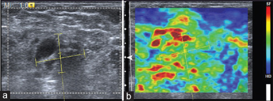
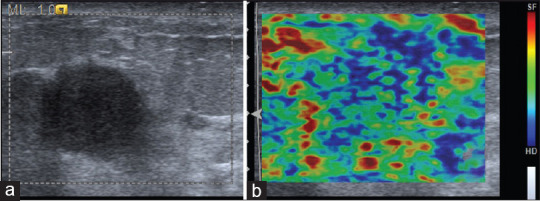
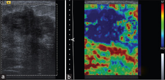
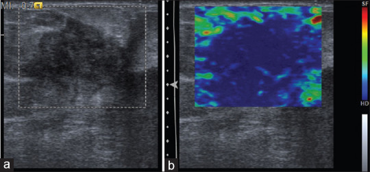
Methods: In this prospective study, 50 palpable breast masses in 50 patients were examined by mammography, B-mode ultrasound (US) and SE. Lesions were categorized using Breast Imaging Reporting and Data System (BIRADS) scoring based on mammographic and sonographic features. Elasticity scores were assessed on a five-point scale based on the distribution of strain, and the lesion size on SE imaging and B-mode (elasticity imaging/B mode [EI/B] ratio) was compared. Findings were correlated with the BIRADS assessment and diagnostic performance of sonoelastography was evaluated taking histopathology as reference standard.
Results: Histopathology revealed 29 (58%) malignant and 21 (42%) benign lesions. Infiltrative ductal carcinoma and fibroadenoma were the most common malignant and benign lesions, respectively. The sensitivity, specificity, positive predictive value, negative predictive value, and accuracy of SE was 100%, 76.1%, 85.2%, 100%, and 90%, respectively. Higher elasticity score was significantly associated with malignant histopathology (P < 0.00001). The mean EI/B ratio for malignant lesions was 1.36 ± 0.24 while that of benign lesions was 1.03 ± 0.30 (P = 0.000).
Conclusion: Real-time SE of the breast, with its superior sensitivity and specificity, could provide improved characterization of benign and malignant breast masses compared with mammography and conventional US. Due to greater diagnostic accuracy, SE can be an effective adjunctive tool to B-mode US in predicting malignancy of breast, as well as in reducing the need for biopsies in benign breast lesions.
Keywords: Breast; elasticity imaging technique; sonoelastography.
Copyright: © 2020 Journal of Medical Ultrasound.
Conflict of interest statement
There are no conflicts of interest.
Figures
References
-
- Zhi H, Ou B, Luo BM, Feng X, Wen YL, Yang HY. Comparison of ultrasound elastography, mammography, and sonography in the diagnosis of solid breast lesions. J Ultrasound Med. 2007;26:807–15. - PubMed
-
- Asteria C, Giovanardi A, Pizzocaro A, Cozzaglio L, Morabito A, Somalvico F, et al. US-elastography in the differential diagnosis of benign and malignant thyroid nodules. Thyroid. 2008;18:523–31. - PubMed
-
- Friedrich-Rust M, Ong MF, Herrmann E, Dries V, Samaras P, Zeuzem S, et al. Real-time elastography for noninvasive assessment of liver fibrosis in chronic viral hepatitis. AJR Am J Roentgenol. 2007;188:758–64. - PubMed
LinkOut - more resources
Full Text Sources