

External Validation of Deep Learning Algorithm for Detecting and Visualizing Femoral Neck Fracture Including Displaced and Non-displaced Fracture on Plain X-ray
- PMID: 34379216
- PMCID: PMC8554912
- DOI: 10.1007/s10278-021-00499-2
External Validation of Deep Learning Algorithm for Detecting and Visualizing Femoral Neck Fracture Including Displaced and Non-displaced Fracture on Plain X-ray
Abstract
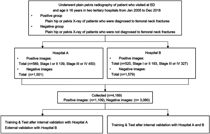
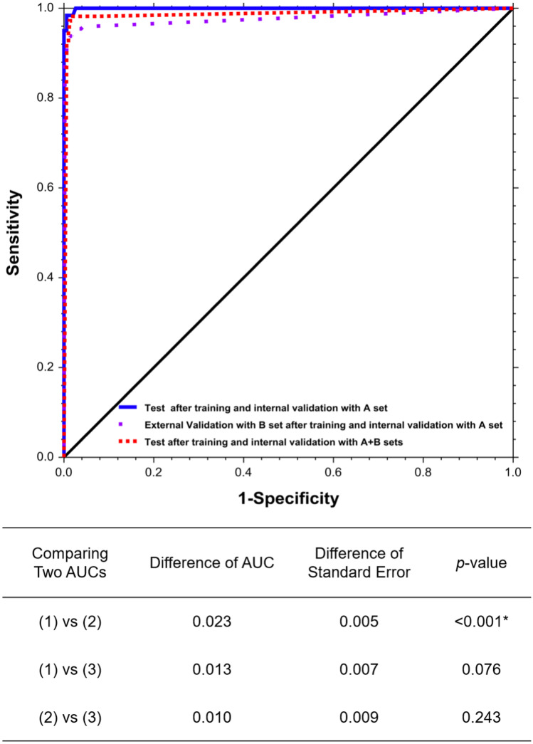
This study aimed to develop a method for detection of femoral neck fracture (FNF) including displaced and non-displaced fractures using convolutional neural network (CNN) with plain X-ray and to validate its use across hospitals through internal and external validation sets. This is a retrospective study using hip and pelvic anteroposterior films for training and detecting femoral neck fracture through residual neural network (ResNet) 18 with convolutional block attention module (CBAM) + + . The study was performed at two tertiary hospitals between February and May 2020 and used data from January 2005 to December 2018. Our primary outcome was favorable performance for diagnosis of femoral neck fracture from negative studies in our dataset. We described the outcomes as area under the receiver operating characteristic curve (AUC), accuracy, Youden index, sensitivity, and specificity. A total of 4,189 images that contained 1,109 positive images (332 non-displaced and 777 displaced) and 3,080 negative images were collected from two hospitals. The test values after training with one hospital dataset were 0.999 AUC, 0.986 accuracy, 0.960 Youden index, and 0.966 sensitivity, and 0.993 specificity. Values of external validation with the other hospital dataset were 0.977, 0.971, 0.920, 0.939, and 0.982, respectively. Values of merged hospital datasets were 0.987, 0.983, 0.960, 0.973, and 0.987, respectively. A CNN algorithm for FNF detection in both displaced and non-displaced fractures using plain X-rays could be used in other hospitals to screen for FNF after training with images from the hospital of interest.
Keywords: AI; Artificial intelligence; Deep learning; Femur; Fracture; Machine learning.
© 2021. Society for Imaging Informatics in Medicine.
Conflict of interest statement
The authors declare no competing interests.
Figures

References
-
- Cummings SR, Rubin SM, Black D. The Future of Hip-Fractures in the United-States - Numbers, Costs, and Potential Effects of Postmenopausal Estrogen. Clin Orthop Relat Res. 1990;252:163–166. - PubMed
Publication types
MeSH terms
LinkOut - more resources
Full Text Sources
Medical
