Review
. 2021 Nov 1;94(1127):20201309.
doi: 10.1259/bjr.20201309. Epub 2021 Aug 11.

Advantages of Colour-Coded Dual-Energy CT Venography in Emergency Neuroimaging

Affiliations
Review

Advantages of Colour-Coded Dual-Energy CT Venography in Emergency Neuroimaging

Camilo A Campo et al. Br J Radiol. .

Abstract

The objective of this Pictorial Review is to describe the use of colour-coded Dual-Energy CT (DECT) to aid in the interpretation of CT Venography (CTV) of the head for emergent indications. We describe a DE CTV acquisition and post-processing technique that can be readily incorporated into clinical workflow. Colour-coded DE CTV may aid the identification and characterization of dural venous sinus abnormalities and other cerebrovascular pathologies, which can improve diagnostic confidence in emergent imaging settings.


Figures

Figure 1.
Figure 1.
Used with permission from Potter et al and RadioGraphics. Three-material decomposition of iodine. A base material line (red line) is defined by the attenuation of cerebrospinal fluid (CSF) and brain parenchyma at low and high kVp. The addition of iodinated contrast (upward blue arrows) increases the attenuation along a characteristic slope, such that everything in the blue-shaded region is attributed to iodine. By projecting back to the base material line along the iodine slope, the attenuation of any voxel (X) in a post-contrast scan can then be decomposed into contributions from the contained iodine and the residual virtual non-contrast (VNC) attenuation.
Figure 2.
Figure 2.
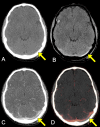
When compared to conventional CTV (A), DECT results can be presented in ways that more clearly differentiate iodine-containing structures from adjacent hyperattenuating structures. (B) Iodine-selective presentation typically displays an iodine map, which highlights the portions of the image that are calculated to contain iodine (i.e., intravenous contrast). (C) DECT post-processing can also subtract the iodine content to create a greyscale virtual non-contrast (VNC) image. (D) Colour-coded overlay presentation superimposes the iodinecontent in colour, on top of the VNC greyscale image, to convey greater anatomic information. Colour-coded iodine content in the dural venous sinuses can be more clearly differentiated from other hyperattenuating materials, such as the adjacent calvarium, when compared to conventional CTV (arrows).
Figure 3.
Figure 3.
Coronal iodine overlay DE CTV image, in which iodine content is colour-coded in orange, superimposed upon grey-scale VNC images. Uniform colour filling the dural venous sinuses makes it easy to assess patency. A prominent right cerebellar developmental venous anomaly (arrow) is also nicely delineated in colour.
Figure 4.
Figure 4.
Patent left transverse sinus stent in a 44-year-old female with history of idiopathic intracranial hypertension presenting to the ED with headache. (A) Conventional CTV demonstrates hyperattenuating contents within the stent (arrow). (B) DE CTV iodine overlay image definitively identifies colour-coded iodinated contrast within the stent (arrow), indicating stent patency rather than stent thrombosis.
Figure 5.
Figure 5.
Left transverse sinus thrombosis in a 21-year-old male with history of unprovoked deep venous thrombosis, presenting to the ED with headache. (A) Conventional CTV demonstrates a normally enhancing right transverse sinus (arrowhead), but slightly diminished attenuation within the left transverse sinus (arrow), suspicious for thrombosis. (B) DE CTV iodine overlay image displays the iodine content in orange, with uniform filling of the right transverse sinus (arrowhead) but a long segment void in the left transverse sinus (arrow), allowing confident diagnosis of sinus thrombosis even in the setting of suboptimal contrast opacification in which venous attenuation is similar to that of thrombus on conventional images.
Figure 6.
Figure 6.
Venous epidural haematoma in a 21-year-old male presenting to the ED after a fall. (A) Non-contrast CT head reveals a non-displaced left occipital bone fracture (not shown) with an underlying hyperattenuating extra axial collection (arrow), best visualised in a soft tissue window to differentiate it from the overlying calvarium. (B) This collection is better demonstrated in a brain window on DECT bone subtraction images that remove the adjacent calvarium (arrow) and is located in the vicinity of the left transverse sinus, raising suspicion for sinus thrombosis or injury. (C) Conventional CTV and (D) colour-coded DECT iodine overlay image demonstrate the collection (arrows) to be a venous epidural haematoma located between the calvarium and the underlying transverse sinus, which is patent but extrinsically compressed.
Figure 7.
Figure 7.
Active parenchymal haemorrhage (“spot sign”) in a 97-year-old female presenting with left-sided neurologic deficits and aphasia. (A) Non-contrast head CT demonstrates a large mixed attenuation right frontal parenchymal haemorrhage with associated leftward midline shift. (B) CTA demonstrates a focus of active bleeding (arrow). (C) The focus of active bleeding increases in size and changes in morphology on conventional CTV (arrow). (D) DE CTV highlights this active extravasation as colour-coded iodine content (arrow), which disappears on the matching VNC image (E). Also note similarity of the VNC image to the true non-contrast image in (A). (F) On the other hand, calcifications within the choroid plexi and the pineal gland (blue arrows) also appear in the colour-coded iodine map, but, unlike iodine, persist on the VNC image (G).

References

    1. Potter CA, Sodickson AD. Dual-Energy CT in emergency neuroimaging: added value and novel applications. Radiographics 2016; 36: 2186–98. doi: 10.1148/rg.2016160069 - DOI - PubMed
    1. Sodickson AD, Keraliya A, Czakowski B, Primak A, Wortman J, Uyeda JW. Dual energy CT in clinical routine: how it works and how it adds value. Emerg Radiol 2021; 28: 103–17. doi: 10.1007/s10140-020-01785-2 - DOI - PubMed
    1. Gupta R, Phan CM, Leidecker C, Brady TJ, Hirsch JA, Nogueira RG, et al. . Evaluation of dual-energy CT for differentiating intracerebral hemorrhage from iodinated contrast material staining. Radiology 2010; 257: 205–11. doi: 10.1148/radiol.10091806 - DOI - PubMed
    1. Phan CM, Yoo AJ, Hirsch JA, Nogueira RG, Gupta R. Differentiation of hemorrhage from iodinated contrast in different intracranial compartments using dual-energy head CT. AJNR Am J Neuroradiol 2012; 33: 1088–94. doi: 10.3174/ajnr.A2909 - DOI - PMC - PubMed
    1. Watanabe Y, Uotani K, Nakazawa T, Higashi M, Yamada N, Hori Y, et al. . Dual-Energy direct bone removal CT angiography for evaluation of intracranial aneurysm or stenosis: comparison with conventional digital subtraction angiography. Eur Radiol 2009; 19: 1019–24. doi: 10.1007/s00330-008-1213-5 - DOI - PubMed