. 2021 Aug 17;118(33):e2103676118.
doi: 10.1073/pnas.2103676118.

A role for Sfrp2 in cardiomyogenesis in vivo

Affiliations

A role for Sfrp2 in cardiomyogenesis in vivo

José A Gomez et al. Proc Natl Acad Sci U S A. .

Abstract

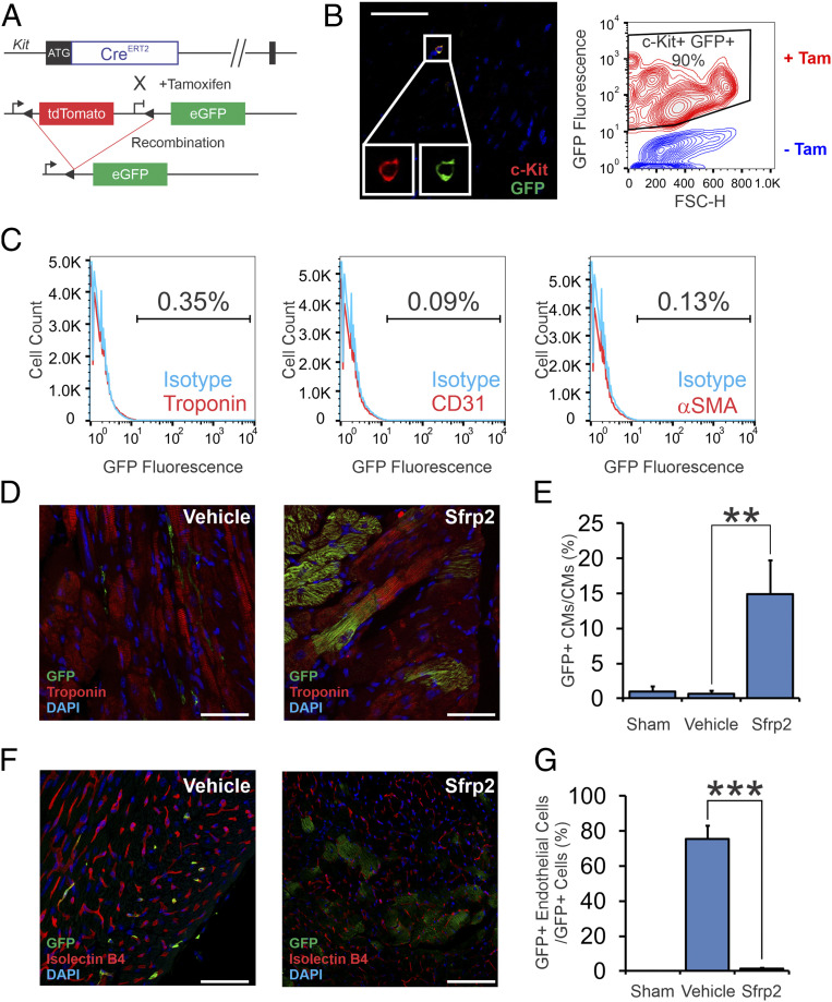
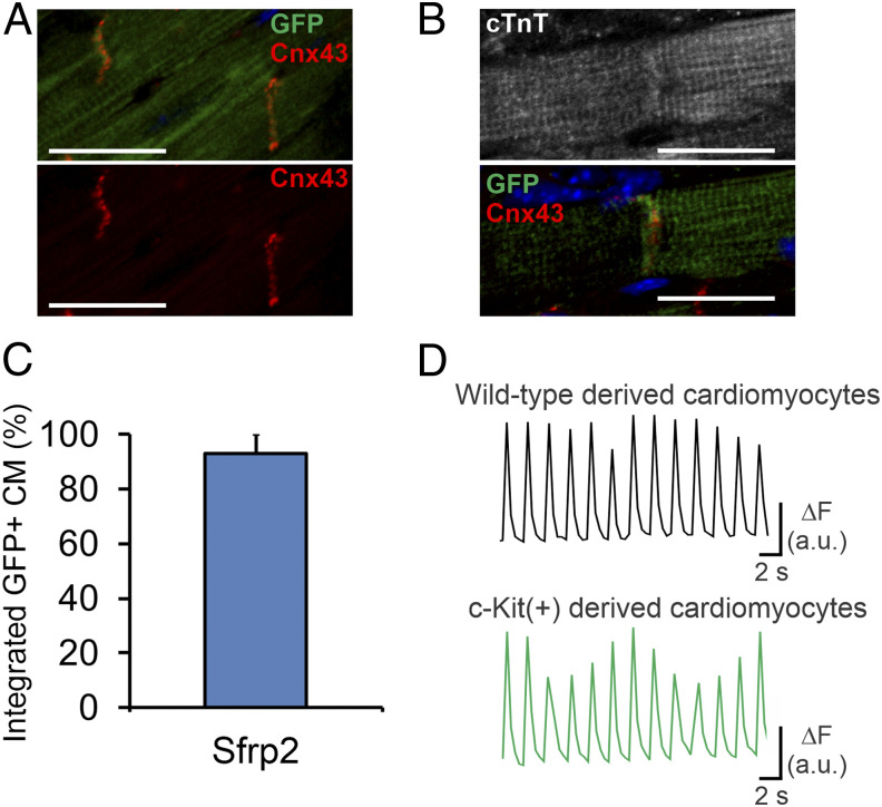
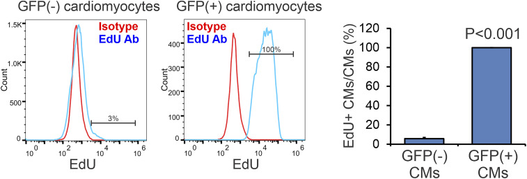
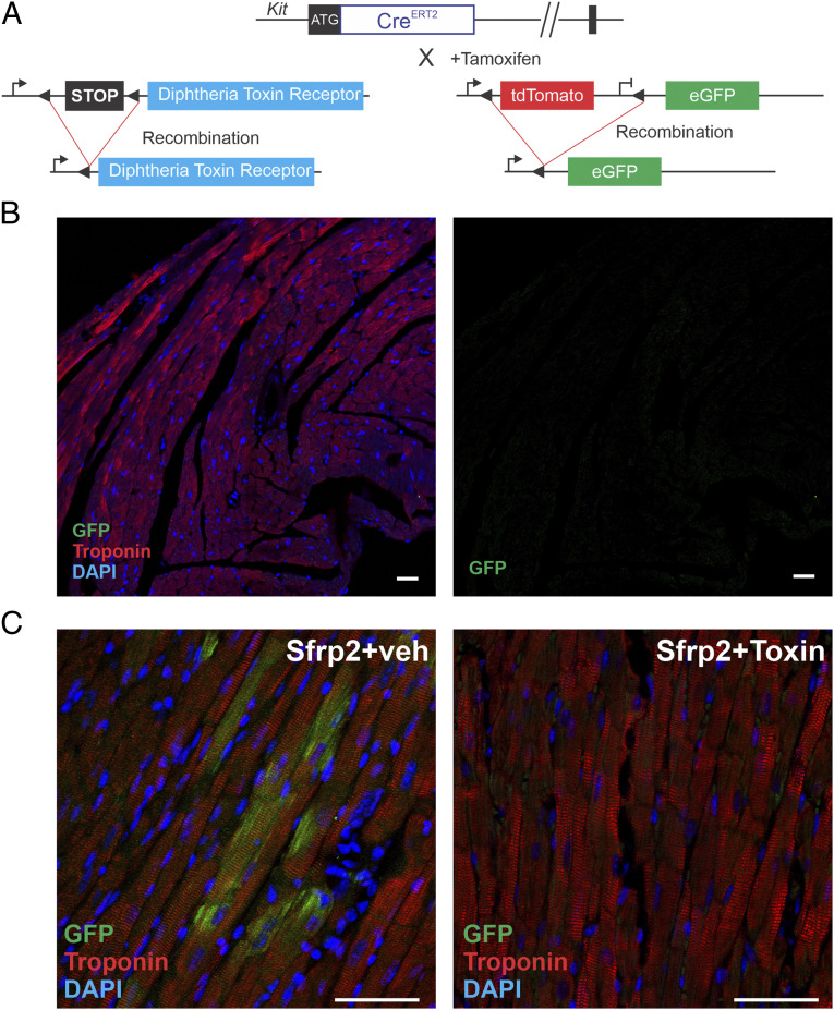
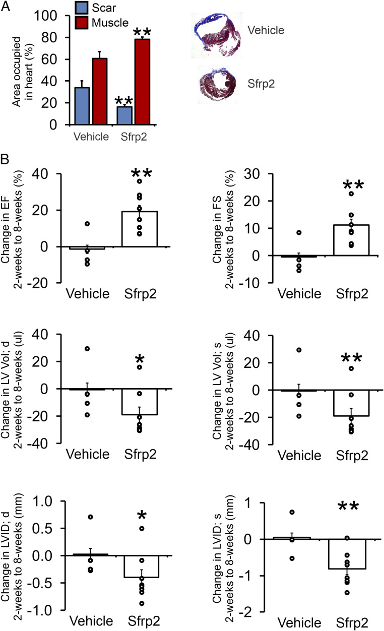
Cardiomyogenesis, the process by which the body generates cardiomyocytes, is poorly understood. We have recently shown that Sfrp2 promotes cardiomyogenesis in vitro. The objective of this study was to determine if Sfrp2 would similarly promote cardiomyogenesis in vivo. To test this hypothesis, we tracked multipotent cKit(+) cells in response to Sfrp2 treatment. In control adult mice, multipotent cKit(+) cells typically differentiated into endothelial cells but not cardiomyocytes. In contrast, Sfrp2 switched the fate of these cells. Following Sfrp2 injection, multipotent cKit(+) cells differentiated solely into cardiomyocytes. Sfrp2-derived cardiomyocytes integrated into the myocardium and exhibited identical physiological properties to preexisting native cardiomyocytes. The ability of Sfrp2 to promote cardiomyogenesis was further supported by tracking EdU-labeled cells. In addition, Sfrp2 did not promote the formation of new cardiomyocytes when the cKit(+) cell population was selectively ablated in vivo using a diphtheria toxin receptor-diphtheria toxin model. Notably, Sfrp2-induced cardiomyogenesis was associated with significant functional improvements in a cardiac injury model. In summary, our study further demonstrates the importance of Sfrp2 in cardiomyogenesis.

Keywords: Wnt signaling pathway; cell differentiation; cell lineage; heart injuries/pathology; myocytes.


Conflict of interest statement

The authors declare no competing interest.

Figures

Fig. 1.
Fig. 1.
Sfrp2 induces cardiomyogenesis in vivo. (A) Description of the cKit(+) lineage tracing mouse. cKitCreERT2/mTeG mice, where the cKit promoter drives the expression of a tamoxifen-inducible Cre, were used. This transgenic mouse that carries a Cre-ERT2 expression cassette inserted into the ATG start codon of the endogenous cKit locus was generated by homologous recombination. Upon tamoxifen treatment, Cre-mediated recombination at the LoxP sites allows expression of eGFP exclusively in cKit(+) cells. (B) Heart tissue was analyzed for eGFP expression in cKit(+) cells by confocal microscopy (Left) and flow cytometry (Right). (Left) Heart tissue from tamoxifen-treated mice was stained with cKit and GFP antibodies and then visualized by confocal microscopy. (Scale bar, 100 μm for representative confocal microscopy images.) Images are from three separate animals. (Right) Heart tissue from vehicle or tamoxifen-treated mice was digested by collagenase, and the resulting single cell suspension was analyzed for cKit and eGFP expression by flow cytometry. CKit(+) cells represented <0.001% of the total population in the heart. Considering the rarity of these cells, a contour plot was used to visualize what is a very small population. (C) Hearts were digested with collagenase. The resulting single cell suspension was analyzed for eGFP expression in cardiomyocytes, endothelial cells, and smooth muscle cells via flow cytometry. (D) Expression of eGFP [cKit(+) cells and cells derived thereof] and the cardiac marker cardiac troponin-T was analyzed by confocal microscopy. Representative confocal images are shown. (Scale bar, 50 μm.) (E) Quantification of D. n = 7 for vehicle-treated group, and n = 10 for Sfrp2-treated group; ANOVA with Bonferroni post hoc tests. Significance shown: **P < 0.01. (F) Cardiac tissue was analyzed for coexpression of GFP and the endothelial cell–specific stain isolectin B4. Representative confocal images are shown. (Scale bar, 50 μm [vehicle] and 100 μm [Sfrp2].) Individual channels are shown in SI Appendix, Fig. S1B. (G) Quantification of F. n = 7 for vehicle-treated group, and n = 10 for Sfrp2-treated group; ANOVA with Bonferroni post hoc tests. Significance shown: ***P<0.001.
Fig. 2.
Fig. 2.
Cardiomyocytes generated via Sfrp2 are physiologically normal and integrate with the myocardium. Following tamoxifen administration to activate the Cre, cKitCreERT2/mTeG mice were subjected to MI. Two days after injury, mice received either vehicle or Sfrp2. Two months after injury, cardiac tissue was analyzed. Sfrp2-derived (eGFP+) cardiomyocytes formed gap junctions with (A) adjacent Sfrp2-derived (eGFP+) cardiomyocytes as well as (B) preexisting (eGFP−) cardiomyocytes, as shown by connexin-43 staining. (Scale bar, 50 μm in A or 25 μm in B.) (C) Quantification of the percentage of Sfrp2-derived (eGFP+) cardiomyocytes, which show apparent integration. n = 7 individual animals. (D) EC coupling in eGFP− and eGFP+ cardiomyocytes. Representative examples of calcium transients obtained from Fura-2–loaded wild-type (eGFP−) and Sfrp2-derived (GFP+) cardiomyocytes during pacing at 0.5 Hz with electric field stimulation.
Fig. 3.
Fig. 3.
Fate-mapping with EdU. Mice were subjected to MI and for the following 10 d received EdU by daily i.p. injection. Six weeks post-MI, eGFP(−) and eGFP(+) cardiomyocytes were analyzed for EdU incorporation. n = 3 for vehicle-treated group, and n = 4 for Sfrp2-treated group; t test, P value shown.
Fig. 4.
Fig. 4.
Genetic ablation prevents Sfrp2-induced cardiomyogenesis. (A) cKitCreERT2/mTmG mice were crossed with a DTR strain to selectively ablate cKit(+) cells in the heart. Mice (cKitCreERT2/mTmG/DTR) were injected with tamoxifen (0.5 mg/mouse) for 14 consecutive days. In the final 7 d of tamoxifen treatment, dipththeria toxin (100 ng/mouse) was injected daily to ablate cKit(+) positive cells. Four days later, treatment mice were subjected to MI. Two days later, mice were injected with Sfrp2 (0.5 µg) or vehicle at the infarct border zone. (B) cKitCreERT2/mTmG/DTR mice were injected with tamoxifen (0.5 mg/mouse) for 14 consecutive days. In the final 7 d of tamoxifen treatment, dipththeria toxin (100 ng/mouse) was injected daily to ablate cKit positive cells. Heart tissue was analyzed for cardiac troponin-T and eGFP expression by confocal microscopy. n = 3. (C) Representative confocal images of 2 mo post-MI hearts. Colocalization of eGFP and the cardiac marker cardiac troponin-T was determined by confocal microscopy. (Scale bar, 50 μm.) n = 5 per group.
Fig. 5.
Fig. 5.
Sfrp2-induced cardiomyogenesis is associated with improved therapeutic outcomes. Mice (cKitCreERT2/mTmG) were injected with tamoxifen (0.5 mg/mouse) for 14 consecutive days. Four days after tamoxifen treatment, mice were subjected to MI. Two days after injury, mice were injected with Sfrp2 (0.5 µg) or PBS at the infarct border zone. (A) Analysis of Masson’s trichrome staining for cardiac muscle (red) and fibrosis (blue). Sections were taken at 0.5 mm and 1 mm below the Sfrp2/vehicle injection point. n = 4 (vehicle) or 8 (Sfrp2); t test, **P < 0.01. Representative Masson’s trichrome–stained cardiac sections are shown. (B) Cardiac function was assessed by echocardiography immediately prior to injury, 2 wk after injury, and finally 8 wk after injury. Raw cardiac function data are provided in SI Appendix, Table S1. Comparisons were made between 2 and 8 wk postinjury in the control and Sfrp2 groups. The graphs show the comparison for each animal (open circle) as well as the average value in each group (bar). n = 4 (vehicle) or 8 (Sfrp2). t tests were carried out between the control and Sfrp2 groups; *P < 0.05, **P < 0.01.

References

    1. Bergmann O., et al. ., Evidence for cardiomyocyte renewal in humans. Science 324, 98–102 (2009). - PMC - PubMed
    1. Jennings R. B., Reimer K. A., The cell biology of acute myocardial ischemia. Annu. Rev. Med. 42, 225–246 (1991). - PubMed
    1. Cleutjens J. P., Blankesteijn W. M., Daemen M. J., Smits J. F., The infarcted myocardium: Simply dead tissue, or a lively target for therapeutic interventions. Cardiovasc. Res. 44, 232–241 (1999). - PubMed
    1. Später D., Hansson E. M., Zangi L., Chien K. R., How to make a cardiomyocyte. Development 141, 4418–4431 (2014). - PubMed
    1. Jeziorowska D., Korniat A., Salem J. E., Fish K., Hulot J. S., Generating patient-specific induced pluripotent stem cells-derived cardiomyocytes for the treatment of cardiac diseases. Expert Opin. Biol. Ther. 15, 1399–1409 (2015). - PubMed

Publication types