. 2021 Aug 11;12(1):4851.
doi: 10.1038/s41467-021-24997-7.

Malaria parasites both repress host CXCL10 and use it as a cue for growth acceleration

Affiliations

Malaria parasites both repress host CXCL10 and use it as a cue for growth acceleration

Yifat Ofir-Birin et al. Nat Commun. .

Abstract

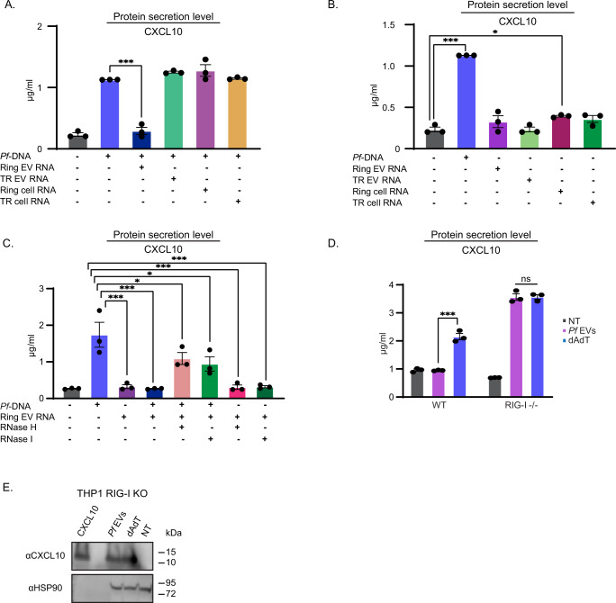
Pathogens are thought to use host molecular cues to control when to initiate life-cycle transitions, but these signals are mostly unknown, particularly for the parasitic disease malaria caused by Plasmodium falciparum. The chemokine CXCL10 is present at high levels in fatal cases of cerebral malaria patients, but is reduced in patients who survive and do not have complications. Here we show a Pf 'decision-sensing-system' controlled by CXCL10 concentration. High CXCL10 expression prompts P. falciparum to initiate a survival strategy via growth acceleration. Remarkably, P. falciparum inhibits CXCL10 synthesis in monocytes by disrupting the association of host ribosomes with CXCL10 transcripts. The underlying inhibition cascade involves RNA cargo delivery into monocytes that triggers RIG-I, which leads to HUR1 binding to an AU-rich domain of the CXCL10 3'UTR. These data indicate that when the parasite can no longer keep CXCL10 at low levels, it can exploit the chemokine as a cue to shift tactics and escape.


Conflict of interest statement

The authors declare no competing interests

Figures

Fig. 1
Fig. 1. Uptake of Pf-derived EVs leads to depletion of chemokine CXCL10 from monocytes.
A RT-PCR analysis of CXCL10, CCL5, IFNA, and IFNB normalized to HPRT1 of THP-1 cells treated with, trophozoite(TR)-derived EVs, ring-derived EVs or not treated (NT) for 6, 12, or 24 h. n = 3 biologically independent experiments, SEM, one-way ANOVA followed by Dunnett’s test, (1 h- CCL5; Ring EVs – NT P <  0.001 *** TR EVs – NT **P = 0.00783. CXCL10; Ring EVs – NT ***P < 0.001. IFNA and IFNB ns not significant. 6 h- CCL5; Ring EVs - ***P < 0.001, TR EVs - NT **P =  0.00514. CXCL10; Ring EVs - NT ***P <  0.001, TR EVs - NT *P = 0.0338. IFNB; Ring EVs - NT ***P < 0.001, TR EVs - NT **P = 0.00597, uRBC EVs - NT **P = 0.00907. IFNA; Ring EVs - NT *P = 0.0229. 24 h- CCL5; Ring EVs - NT ***P <  0.001. CXCL10; Ring EVs - NT ***P <  0.001, TR EVs - NT *P = 0.0417. IFNB; Ring EVs - NT ***P <  0.001. IFNA; Ring EVs - NT ***P < 0.001). B THP-1 cells were incubated with Pf-ring-stage-, Pf-trophozoite-stage- or uRBC-derived vesicles for 1, 6, and 24 h. An ELISA assay was performed on the cells’ media to detect secreted CCL5 and CXCL10. HEK blue IFNα/β assay was performed. n = 3 biologically independent experiments, SEM, one-way ANOVA followed by Dunnett’s test, CXCL10; ns. CCL5; 1-h-ns, 6 h- Ring EVs - uRBC EVs ***P <  1e-10, 24 h- Ring EVs - uRBC EVs ***P =  1.03e-10. IFNα/β- 1 h- Ring EVs - uRBC ***P = 4.17e-06, TR EVs - uRBC EVs *P = 0.0138. 6 h- Ring EVs - uRBC EVs ***P = 1.24e-08, TR EVs - uRBC EVs ***P = 0.000847, 24 h- Ring EVs - uRBC EVs ***P = 9.66e-08, TR EVs - uRBC EVs ***P = 0.000459). C Western Blot assay of CXCL10 and HSP90 (loading control) was performed on THP-1 cell lysate. THP-1 cells were incubated with Pf-ring-stage-derived EVs and then transfected with poly(dA:dT) or not treated (NT) for 16 h before harvesting. Results are representative of at least three independent biological replicates. D THP-1 cells were incubated with Pf-ring-stage-derived EVs and then transfected with poly(dA:dT) or NT for 16, 20 or 24 h. An ELISA assay was performed on the cell lysate for CXCL10. n = 3 biologically independent experiments, SEM, one-way ANOVA followed by Dunnett’s test, (16 h dAdT - NT ***P = 1.68e-05. 20 hours dAdT - NT ***P = 6.28e-07. 24 h dAdT - NT ***P = 2.44e-06). E Western Blot analysis of THP-1 cell lysate. THP-1 cells were incubated with Pf-ring-stage-derived EVs, transfected with poly(dA:dT) and treated with Pf-ring-stage-derived EVs for 1 h before being transfected with poly(dA:dT). The cells were harvested 16, 20, and 24 h post treatment. Antibodies were used against CXCL10 and HSP90 (loading control). NT not treated. Results are representative of at least three independent biological replicates. F ELISA assay for CXCL10 was performed on cell lysate. THP-1 cells were incubated with Pf-ring-stage-derived EVs, transfected with poly(dA:dT), and then treated with Pf-ring-stage-derived EVs for 1 h before being transfected with poly(dA:dT). Cells were harvested 16, 20, and 24 h post treatment NT-not treated. n = 3 biologically independent experiments, SEM. One-way ANOVA followed by Dunnett’s test, (16 h- dAdT - NT ***P <  1e-04, Pf EVs+dAdT - NT ***P = 0.000245. 20 h- dAdT - NT ***P <  0.001, Pf EVs+dAdT - NT **P = 0.00475. 24 h- dAdT - NT ***P < 0.001, Pf EVs+dAdT - NT P <  0.001 ***). G ELISA assay for detection of CXCL10 in PBMC-derived monocytes. CD14+ primary cells (monocytes) isolated from three naive healthy donors. The cells were incubated with Pf-ring-stage-derived EVs or transfected with poly(dA:dT) and then analyzed by ELISA. n = 3 biologically independent experiments, SEM, one-way ANOVA followed by Dunnett’s test, (dAdT - NT ***P =  1.29e-07) Source data are provided as a Source Data file. .
Fig. 2
Fig. 2. Pf-derived EVs inhibit the translation of CXCL10.
A Polysomal profiling of THP-1 cells 14 h post transfection with poly(dA:dT) (blue), Pf –derived EVs (purple), or not treated (NT) (black). One representative repeat out of five biological replicates is shown. B. THP-1 cells were incubated with Pf-ring-stage-derived EVs or transfected with poly(dA:dT) for 14 h or NT. Polysomal profiling was performed on all samples and RNA was isolated from heavy, light and free fractions. Real-time PCR for CXCL10 was performed on all the polysome fractions. n = 3 biologically independent experiments, NT (not treated) THP-1 cells were used as control. One-way ANOVA followed by Dunnett’s test for the heavy fractions (dAdT - NT ***P = 1.64e-05, Pf EVs - NT *P = 0.0368). C. THP-1 cells were incubated with Pf-ring-stage-derived EVs, transfected with poly(dA:dT) for 14 h or NT. Ribosomal profiling was performed on all samples and RNA was isolated from heavy, light and free fractions. Real-time PCR for IFNB was performed on all the polysome fractions. n = 3 biologically independent experiments. One-way ANOVA followed by Dunnett’s test for the heavy fractions (dAdT - NT ***P = 3.23e-05, Pf EVs - NT ***P = 0.000543). Source data are provided as a Source Data file.
Fig. 3
Fig. 3. Selected RNA cargo mediates CXCL10 translation inhibition via the RIG-I receptor.
A THP-1 cells were transfected with RNA purified from: Pf ring stage, trophozoite stage, Pf-ring-stage-derived EVs or EVs derived from Pf-trophozoite-(TR) stage. In addition, all the cells were transfected with Pf gDNA. An ELISA was performed on the cell media for CXCL10 16 h post transfection. n = 3 biologically independent experiments, SEM, one-way ANOVA followed by Dunnett’s test (Pf-DNA + Ring EVs RNA - Pf-DNA ***P <  0.001). NT not treated. B THP-1 cells were transfected with Pf gDNA, NT, or RNA purified from: Pf ring stage, trophozoite stage, Pf-ring-stage-derived EVs, or Pf-trophozoite-stage-derived EVs. An ELISA was performed on the cell media for CXCL10 16 h post transfection. n = 3 biologically independent experiments, SEM, one-way ANOVA followed by Dunnett’s test, (Pf-DNA - NT ***P <  0.001, Ring-cell RNA - NT *P = 0.0405). C THP-1 cells were transfected with RNA purified from: Pf-derived EVs, Pf-derived-EVs pretreated with RNase H and Pf-derived EVs pretreated with RNase I. In addition, the cells were transfected with Pf gDNA or NT. An ELISA was performed on the cell media for CXCL10 16 h post transfection. n = 3 biologically independent experiments, SEM, one-way ANOVA followed by Dunnett’s test, (NT- Pf-DNA ***P <  0.001, Pf-DNA + Ring EVs RNA - Pf-DNA ***P < 0.001, Pf-DNA + RNase H - Pf-DNA *P = 0.0439, Pf-DNA + RNase I - Pf-DNA *P = 0.0112, Ring EVs-RNA - Pf-DNA ***P <  0.001, RNase H - Pf-DNA ***P < 0.001, RNase I - Pf-DNA ***P < 0.001). D THP-1 RIG-I KO cells were incubated with EVs harvested from ring-stage Pf (iRBCs) and then transfected with poly(dA:dT) or NT for 24 h. An ELISA assay for detecting CXCL10 was performed on cell lysates. n = 3 biologically independent experiments, SEM, one-way ANOVA followed by Dunnett’s test, (THP-1 WT- Pf EVs-dAdT ***P = 0.0000428). E Western Blot assay of CXCL10 and HSP90 (loading control) were performed on THP-1 RIG-I KO cells. The THP-1 cells were incubated with Pf-ring-stage-derived EVs, transfected with poly(dA:dT) or not treated (NT) 16 h before harvesting. Results are representative of at least three independent biological replicates. Source data are provided as a Source Data file.
Fig. 4
Fig. 4. HUR-1 binds to AU- rich domain in the 3′UTR of the CXCL10 transcript and is involved in translation inhibition.
A Schematic illustration of CXCL10 mRNA. Orange squares, blue rectangles, and gray arrows represent the UTRs, exons, and introns of the CXCL10 mRNA, respectively. The green circles illustrate the AU-rich domains predicted by http://arescore.dkfz.de/ analysis. B THP-1 cells were incubated with Pf-derived EVs 5 or 16 h (H) or not treated before the transfection with the CXCL10-3′UTR-Luc plasmid. 24 h post transfection, the cell media was collected, luciferase levels were measured and normalized to SEAP levels. n = 3 biologically independent experiments, SEM, one-way ANOVA followed by Dunnett’s test, (3UTR+Pf EVs 16 h - ***P <  1e-04, 3UTR + Pf EVs 5 h - 3UTR ***P <  1e-04). C THP-1 RIG-I KO cells were incubated with Pf-ring-stage-derived EVs for 5 or 16 h (H) or not treated (NT) before their transfection with the CXCL10-3′UTR-Luc plasmid. 24 hours post transfection, the cell media was collected and luciferase levels were measured and normalized to the SEAP levels. n = 3 biologically independent experiments, SEM, one-way ANOVA followed by Dunnett’s test (ns). D Schematic illustration of the three sub-domains of CXCL10 3′UTR and the cloned deletions ΔI, ΔII, and ΔIII. E THP-1 cells were treated with Pf -derived EVs for 16 h prior to transfection with CXCL10 3′UTR-fused-reporter, ΔI, ΔII, ΔIII plasmids (NT-not treated cells). 24 hours post-transfection the cells media was collected, Luciferase levels were measured and normalized to the SEAP levels. n = 3 biologically independent experiments, SEM, one-way ANOVA followed by Dunnett’s test, (Pf EVs- 3UTR ΔII - 3UTR ***P <  1e-06). F THP-1 cells were transfected with siRNA for HUR1 or with non-targeting siRNA pool (NR). 48 hours post siRNA transfection, cells were treated with Pf-derived EVs, with poly(dA:dT) and following 24 h post treatments, secreted CXCL10 in the cells media was measured by an ELISA assay, n = 4 biologically independent experiments, SEM, one-way ANOVA followed by Dunnett’s test, (NT- Pf-DNA - NT ***P = 1.14e-06, NR- Pf-DNA - NT ***P = 5.14e-07, siHUR1- Pf-DNA - NT ***P = 2.59e-05, Pf-EVs - NT **P = 0.00907). NT (not treated). G THP-1 cells were transfected with siRNA for HUR1 or with non-targeting siRNA pool (NR). 48 hours post siRNA transfection, cells were treated with Pf-derived EVs, with poly(dA:dT) and following 24 h post treatments, HUR1 reduction and CXCL10 translation was verified by Western Blot analysis. HSP90 used as a loading control. NT (not treated). Results are representative of at least three independent biological replicates. H THP-1 cells were treated with Pf -derived EVs or transfected with poly(dA:dT). Six hours post treatment, cells were discarded and native RIP assay was performed using anti-HUR1 antibody or anti-HSP70 as negative control. Real-time PCR analysis for CXCL10, CCL5 (as negative control) and c-Myc (as positive control) was performed on the input (pull down) of the RIP samples, n = 3 biologically independent experiments, SEM, one-way ANOVA followed by Dunnett’s test, (Pf EVs -*P = 0.0427). NT (not treated). Source data are provided as a Source Data file.
Fig. 5
Fig. 5. The chemokine CXCL10 promotes parasite growth.
A NF54 Pf parasites were cultured with media containing: (1) complete media [lacking CXCL10] (Black, RPMI); (2) media harvested from untreated THP-1 cells [lacking CXCL10] (Green, CM-NT); (3) media harvested from untreated THP-1 cells with supplemental 25 ng/ml CXCL10 [containing CXCL10] (gray, CM-NT+CXCL10); (4) media harvested from THP-1 cells pretreated with Pf-ring-stage-derived EVs [lacking CXCL10] (pink, CM-Pf-EVs); 5) Media harvested from THP-1 cells pretreated with Pf-ring-stage-derived EVs and supplemental 25 ng/ml CXCL10 [containing CXCL10] (red, CM-Pf EVs+CXCL10); or (6) Media harvested from THP-1 cells transfected with Pf gDNA [containing CXCL10] (blue, CM-Pf DNA). Parasitemia levels were measured every second day using flow cytometry. Graph shows relative parasitemia level. SEM, n = 5 biologically independent experiments, one-way ANOVA followed by Dunnett’s test, (1 day- CM-NT - RPMI *P = 0.0435, 3 day- CM-NT + CXCL10 - RPMI **P = 0.00480, CM-Pf DNA - RPMI ***P <  0.001, CM-Pf EVs + CXCL10 - RPMI **P = 0.00489, 5 day- CM-NT - RPMI **P = 0.00502, CM-NT + CXCL10 - RPMI ***P < 0.001, CM-Pf DNA - RPMI ***P < 0.001, CM-Pf EVs + CXCL 10 - RPMI ***P < 0.001). The data was analyzed using Diva v. 8.0.1 software (BD). The gating strategy is demonstrated in Supplementary Figure 27. B Representative Giemsa smears from a growth assay (A). Size bar 10 µm. Results are representative of at least three independent biological replicates. C Parasitemia counting using Giemsa smears from day 5 of the parasite culture (A). n = 3 biologically independent experiments, SEM, one-way ANOVA followed by Dunnett’s test, (CM-NT - RPMI *P = 0.0373, CM-NT + CXCL10 - RPMI ***P < 0.001, CM-Pf DNA - RPMI ***P < 0.001, CM-Pf EVs - RPMI *P = 0.0351, CM-Pf EVs + CXCL 1 0 – RPMI ***P < 0.001). Source data are provided as a Source Data file.
Fig. 6
Fig. 6. proposed two-phase mechanism of CXCL10 regulation.
A ‘decision sensing system’: A In non-complicated malaria (upper panel) the Pf-iRBCs repress CXCL10 expression in host monocytes via transferring of RNA cargo encapsulated in parasite-derived EVs. The RNA cargo is then recognized by the host RNA receptor, RIG-I, leading to a cascade of events that eventually blocks the association of the ribosomes to CXCL10 transcript in a process mediated by its 3′UTR and binding of HUR1. B However, in complicated malaria (such as CM) when CXCL10 level are higher, the chemokine is sensed back as an ‘alert facilitator’, prompting parasitic proliferation, possibly allowing an escape action (bottom panel). Other host immune cells produce and secrete CXCL10.

References

    1. Rieke B. Overview of the worldwide Handling with the Disease in Prevention, Diagnostics and Therapy WHO World Malaria Report 2017. Flugmed. Tropenme. 2018;25:5–5.
    1. Verlinden BK, Louw A, Birkholtz LM. Resisting resistance: is there a solution for malaria? Expert Opin. Drug Discov. 2016;11:395–406. doi: 10.1517/17460441.2016.1154037. - DOI - PubMed
    1. Dunst J, Kamena F, Matuschewski K. Cytokines and chemokines in cerebral malaria pathogenesis. Front. Cell. Infect. Microbiol. 2017;7:324. doi: 10.3389/fcimb.2017.00324. - DOI - PMC - PubMed
    1. Jain V, et al. Plasma IP-10, apoptotic and angiogenic factors associated with fatal cerebral malaria in India. Malar. J. 2008;7:83. doi: 10.1186/1475-2875-7-83. - DOI - PMC - PubMed
    1. Wilson NO, et al. CXCL4 and CXCL10 predict risk of fatal cerebral malaria. Dis. Markers. 2011;30:39–49. doi: 10.1155/2011/828256. - DOI - PMC - PubMed

Publication types

MeSH terms