. 2021 Sep;28(9):740-746.
doi: 10.1038/s41594-021-00651-0. Epub 2021 Aug 11.

Mechanism of molnupiravir-induced SARS-CoV-2 mutagenesis

Affiliations

Mechanism of molnupiravir-induced SARS-CoV-2 mutagenesis

Florian Kabinger et al. Nat Struct Mol Biol. 2021 Sep.

Abstract

Molnupiravir is an orally available antiviral drug candidate currently in phase III trials for the treatment of patients with COVID-19. Molnupiravir increases the frequency of viral RNA mutations and impairs SARS-CoV-2 replication in animal models and in humans. Here, we establish the molecular mechanisms underlying molnupiravir-induced RNA mutagenesis by the viral RNA-dependent RNA polymerase (RdRp). Biochemical assays show that the RdRp uses the active form of molnupiravir, β-D-N4-hydroxycytidine (NHC) triphosphate, as a substrate instead of cytidine triphosphate or uridine triphosphate. When the RdRp uses the resulting RNA as a template, NHC directs incorporation of either G or A, leading to mutated RNA products. Structural analysis of RdRp-RNA complexes that contain mutagenesis products shows that NHC can form stable base pairs with either G or A in the RdRp active center, explaining how the polymerase escapes proofreading and synthesizes mutated RNA. This two-step mutagenesis mechanism probably applies to various viral polymerases and can explain the broad-spectrum antiviral activity of molnupiravir.


Conflict of interest statement

The authors declare no competing interests.

Figures

Fig. 1
Fig. 1. RdRp incorporates NHC opposite G and A in the template.
a, Chemical structure of molnupiravir. b, Chemical structure of NHC triphosphate (MTP). c, The RNA template–product duplex. The direction of RNA extension is shown. The color of the depicted circles indicates the experimental design: blue, RNA template strand; dark blue, +1 templating nucleotide; red, RNA product strand; gray, NTP substrate; orange, MTP. The 5′ end of the RNA product contains a FAM fluorescent label. C* at the 3′ end of the template indicates dideoxy-C (ddC). d, NHC monophosphate is incorporated into growing RNA instead of C or U when G or A are present in the template +1 position. e, Quantification of nucleotide incorporation efficiency relative to the cognate NTP (dark gray) after triplicate measurements. Non-cognate NTPs and MTP are depicted in light gray and orange, respectively. Individual data points and boxes represent mean ± s.d. f, Quantification of time-dependent M incorporation opposite a templating G residue after triplicate measurements. Incorporation efficiency is calculated relative to cognate C incorporation. Data are mean ± s.d. g, Quantification of time-dependent M incorporation opposite a templating A residue after triplicate measurements. Incorporation efficiency is calculated relative to cognate U incorporation. Data are mean ± s.d. An uncropped gel image for d and data behind the graphs in eg are available as source data. Source data
Fig. 2
Fig. 2. NHC incorporation does not stall SARS-CoV-2 RdRp.
a, The RNA template–product duplex (as in Fig. 1c) allows for RNA extension by four nucleotides. The direction of RNA extension is indicated. The 5′ end of the RNA product contains a FAM fluorescent label. C* at the 3′ end of the template indicates dideoxy-C (ddC). b, RNA elongation to the end of the template in a is possible when MTP replaces either CTP or UTP in the presence of adenosine triphosphate (ATP). The experiment was performed once. c, The RNA template–product hairpin duplex allows for RNA extension by 11 nucleotides. d, RNA elongation stalls at the expected positions when the cognate NTP is withheld from the reaction. Extension to the end of the template is possible when MTP replaces either CTP or UTP in the presence of other substrate NTPs, showing that incorporation of M does not prevent RNA extension. Note that more efficient RNA extension is seen at higher NTP/MTP concentrations, and also for MTP replacing UTP (not shown). The experiment was performed once. Uncropped gel images for b and d are available as source data. Source data
Fig. 3
Fig. 3. NHC can direct incorporation of G and A into RNA.
a, Scheme of synthesis of RNA containing NHC monophosphate (M) at a defined position. 5′-O-DMT-2′-O-TOM-protected N4-hydroxycytidine phosphoramidite (M-PA) used for solid-phase synthesis of M-containing RNA (M-RNA). b, Analysis of M-containing RNA by denaturing HPLC confirms the homogeneity of the synthetic RNA (top). HR-ESI-MS analysis proves the presence of NHC and absence of unmodified RNA (bottom). c, The RNA template–product scaffold with M in template position +1, where it is used by the RdRp to direct binding of the incoming NTP substrate. The 5′ end of the RNA product contains a FAM fluorescent label. C* at the 3′ end of the template indicates dideoxy-C (ddC). d, When present at position +1 of the template strand, M can direct the incorporation of G or A into nascent RNA, but not C or U. e, Quantification of the experiment in d after triplicate measurements. Incorporation efficiencies are calculated relative to C incorporation opposite templating G. Individual data points and error bars represent mean ± s.d. An uncropped gel image for d and data behind the graph in e are available as source data. Source data
Fig. 4
Fig. 4. Structures of RdRp–RNA product complexes after NHC-induced mutagenesis.
a, Overview of RdRp–RNA structure with an M residue (orange) at position −1 in the RNA template strand. RdRp subunits nsp7, nsp8 and nsp12 are in dark blue, green and gray, respectively. The RNA template and product are in blue and red, respectively. The active site is indicated by a magenta sphere. Depicted is the structure containing the M-A base pair. b, RNA duplex containing the M-A base pair in the RdRp active center. The +1 position (templating nucleotide, NTP substrate site) and the −1 position (post-translocation position of the nascent base pair) are indicated. c, RNA duplex containing the M-G base pair in the RdRp active center. d, Cryo-EM density for the nascent M-A (top) and M-G (bottom) base pairs in position −1, viewed along the RNA duplex axis in the direction of RNA translocation. e, M-A (top) and M-G (bottom) base pairing relies on different tautomeric forms of NHC, as predicted.
Fig. 5
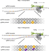
Fig. 5. Two-step model of molnupiravir-induced RNA mutagenesis.
In the presence of NTPs and MTP, M nucleotides can be incorporated by SARS-CoV-2 RdRp instead of C or U into the negative-strand genomic (−gRNA) or subgenomic RNA (−sgRNA) during copying of the positive-strand genomic RNA template (+gRNA). The obtained M-containing negative-strand RNAs can then be used as a template for the production of mutagenized +gRNA and positive-strand subgenomic mRNA (+sgmRNA). These RNA products are predicted to be mutated and not to support formation of functional viruses. RNA of random sequence is shown, with M and mutated residues indicated as orange and violet letters, respectively.
Extended Data Fig. 1
Extended Data Fig. 1. Synthesis of NHC-phosphoramidite 3 (M-PA).
5’-O-DMT-2’-O-TOM-O4-chlorophenyluridine (A) was synthesized from uridine as previously reported (L. Buttner, J. Seikowski, K. Wawrzyniak, A. Ochmann, C. Hobartner, Synthesis of spin-labeled riboswitch RNAs using convertible nucleosides and DNA-catalyzed RNA ligation. Bioorg Med Chem 21, 6171-6180 (2013)). The chlorophenol group was displaced by hydroxylamine to give new compound 1. After selective benzoyl protection at N4 with benzoic anhydride, compound 2 was converted to the phosphoramidite M-PA (3) using 2-cyanoethyl-N,N,N’,N’-tetraisopropylphosphorodiamidite and 4,5-dicyano-imidazol (DCI) in analogy to a previous report (J. Lu, L. Nan-Sheng, J. A. Piccirilli, Efficient Synthesis of N 4-Methyl- and N 4-Hydroxycytidine Phosphoramidites. Synthesis 16, 2708-2712 (2010)). DMT-Cl = 4,4’-dimethoxy-trityl chloride, TOM-Cl = triisopropylsilyloxymethyl chloride, DBU = 1,8-diazabicyclo [5.4.0]undec-7-ene, DMAP = 4-(N,N-dimethylamino)- pyridine.
Extended Data Fig. 2
Extended Data Fig. 2. Melting curves for RNA duplexes containing M–G or M–A base pairs.
UV thermal melting monitored at 260 nm for 20 µM duplexes (11 bp, 4 nt singlestranded overhang) in 100 mM NaCl, 10 mM Na-phosphate buffer pH 7.0. black: unmodified duplex C–G (Tm = 64.7 °C), red terminal M–G (Tm = 61.2 °C): blue: terminal M–A base pair (Tm = 60.6 °C). See also Supplementary Table 2.
Extended Data Fig. 3
Extended Data Fig. 3. Protein preparation and RNA scaffold for structural studies.
a, RNA scaffold was obtained by annealing a short M-containing oligonucleotide to a hairpin RNA duplex. b, SDS-PAGE of purified RdRp-RNA complexes used for cryo EM. Purified proteins were run on 4-12 % Bis-Tris SDS-PAGE gels in 1x MOPS buffer and stained with Coomassie Blue. M-A corresponds to the RdRp complex with RNA scaffold, where M in template base pairs to A. M-G corresponds to the RdRp complex with RNA scaffold, where M in template base pairs to G. L: PageRuler Prestained Protein Ladder (Thermo Scientific). The experiment was performed once, source data are provided as a Source Data file. Source data
Extended Data Fig. 4
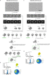
Extended Data Fig. 4. Cryo-EM data processing trees and quality of reconstructions.
a, Cryo-EM data processing tree for M–A bp-containing RdRp-RNA structure. Scale bar, 100 nm. b, local resolution, FSC plot, angular distribution and directional FSC calculated according to Tan et al., Nature Methods 14, 793–796 (2017). Sphericity of M-A containing structure is 0.976; c, Cryo-EM data processing tree for M–G bp-containing RdRp-RNA structure. Scale bar, 100 nm. d, Local resolution, FSC plot, angular distribution and directional FSC calculated according to Tan et al., Nature Methods 14, 793–796 (2017). Sphericity of M-G containing structure is 0.966.

Comment in

References

    1. Dolgin E. The race for antiviral drugs to beat COVID—and the next pandemic. Nature. 2021;592:340–343. doi: 10.1038/d41586-021-00958-4. - DOI - PubMed
    1. Hilgenfeld R, Peiris M. From SARS to MERS: 10 years of research on highly pathogenic human coronaviruses. Antivir. Res. 2013;100:286–295. doi: 10.1016/j.antiviral.2013.08.015. - DOI - PMC - PubMed
    1. Snijder EJ, Decroly E, Ziebuhr J. The nonstructural proteins directing coronavirus RNA synthesis and processing. Adv. Virus Res. 2016;96:59–126. doi: 10.1016/bs.aivir.2016.08.008. - DOI - PMC - PubMed
    1. Posthuma CC, Te Velthuis AJW, Snijder EJ. Nidovirus RNA polymerases: complex enzymes handling exceptional RNA genomes. Virus Res. 2017;234:58–73. doi: 10.1016/j.virusres.2017.01.023. - DOI - PMC - PubMed
    1. Romano M, Ruggiero A, Squeglia F, Maga G, Berisio R. A structural view of SARS-CoV-2 RNA replication machinery: RNA synthesis, proofreading and final capping. Cells. 2020;9:1267. doi: 10.3390/cells9051267. - DOI - PMC - PubMed

Publication types

MeSH terms