

The Roles of Kidney-Resident ILC2 in Renal Inflammation and Fibrosis
- PMID: 34381446
- PMCID: PMC8350317
- DOI: 10.3389/fimmu.2021.688647
The Roles of Kidney-Resident ILC2 in Renal Inflammation and Fibrosis
Abstract
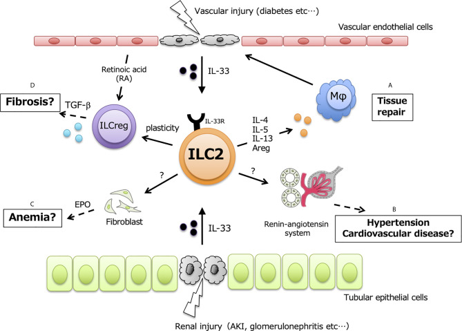
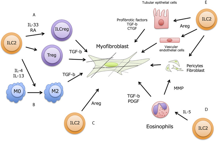
Innate lymphoid cells (ILCs) are a recently discovered lymphocyte population with high cytokine productive capacity. Type-2 ILCs (ILC2s) are the most studied, and they exert a rapid type-2 immune response to eliminate helminth infections. Massive and sustainable ILC2 activation induces allergic tissue inflammation, so it is important to maintain correct ILC2 activity for immune homeostasis. The ILC2-activating cytokine IL-33 is released from epithelial cells upon tissue damage, and it is upregulated in various kidney disease mouse models and in kidney disease patients. Various kidney diseases eventually lead to renal fibrosis, which is a common pathway leading to end-stage renal disease and is a chronic kidney disease symptom. The progression of renal fibrosis is affected by the innate immune system, including renal-resident ILC2s; however, the roles of ILC2s in renal fibrosis are not well understood. In this review, we summarize renal ILC2 function and characterization in various kidney diseases and highlight the known and potential contributions of ILC2s to kidney fibrosis.
Keywords: CKD - chronic kidney disease; IL-33; ILC2; ILCreg; renal fibrosis.
Copyright © 2021 Nagashima and Iyoda.
Conflict of interest statement
The authors declare that the research was conducted in the absence of any commercial or financial relationships that could be construed as a potential conflict of interest.
Figures
Similar articles
-
The IL-33/ST2/ILC2 pathway in kidney disease: balancing inflammation, fibrosis, and repair.Am J Physiol Cell Physiol. 2025 Sep 1;329(3):C718-C725. doi: 10.1152/ajpcell.00405.2025. Epub 2025 Jul 24. Am J Physiol Cell Physiol. 2025. PMID: 40707033 Review.
-
Group2 innate lymphoid cells ameliorate renal fibrosis and dysfunction associated with adenine-induced CKD.Cell Immunol. 2024 Jul-Aug;401-402:104828. doi: 10.1016/j.cellimm.2024.104828. Epub 2024 May 12. Cell Immunol. 2024. PMID: 38759328
-
IL-33 attenuates renal fibrosis via group2 innate lymphoid cells.Cytokine. 2022 Sep;157:155963. doi: 10.1016/j.cyto.2022.155963. Epub 2022 Jul 19. Cytokine. 2022. PMID: 35868116
-
Macrophages in kidney injury, inflammation, and fibrosis.Physiology (Bethesda). 2015 May;30(3):183-94. doi: 10.1152/physiol.00046.2014. Physiology (Bethesda). 2015. PMID: 25933819 Review.
-
Group 2 innate lymphoid cells contribute to IL-33-mediated alleviation of cardiac fibrosis.Theranostics. 2021 Jan 1;11(6):2594-2611. doi: 10.7150/thno.51648. eCollection 2021. Theranostics. 2021. PMID: 33456562 Free PMC article.
Cited by
-
Innate immune cellular therapeutics in transplantation.Front Transplant. 2023;2:1067512. doi: 10.3389/frtra.2023.1067512. Epub 2023 Mar 31. Front Transplant. 2023. PMID: 37994308 Free PMC article.
-
Renal IL-23-Dependent Type 3 Innate Lymphoid Cells Link Crystal-induced Intrarenal Inflammasome Activation with Kidney Fibrosis.J Immunol. 2024 Sep 15;213(6):865-875. doi: 10.4049/jimmunol.2400041. J Immunol. 2024. PMID: 39072698 Free PMC article.
-
A novel comprehensive strategy for research on wine-processed mechanism of corni fructus guided by variation in the chemical components and network analysis.BMC Complement Med Ther. 2025 Aug 21;25(1):312. doi: 10.1186/s12906-025-05046-y. BMC Complement Med Ther. 2025. PMID: 40841951 Free PMC article.
-
A Deep View of the Biological Property of Interleukin-33 and Its Dysfunction in the Gut.Int J Mol Sci. 2023 Aug 31;24(17):13504. doi: 10.3390/ijms241713504. Int J Mol Sci. 2023. PMID: 37686309 Free PMC article. Review.
-
Human umbilical cord mesenchymal stem cell-derived exosomal miR-335-5p attenuates the inflammation and tubular epithelial-myofibroblast transdifferentiation of renal tubular epithelial cells by reducing ADAM19 protein levels.Stem Cell Res Ther. 2022 Jul 28;13(1):373. doi: 10.1186/s13287-022-03071-z. Stem Cell Res Ther. 2022. PMID: 35902972 Free PMC article.
References
-
- Cruz-Solbes AS, Youker K. Epithelial to Mesenchymal Transition (EMT) and Endothelial to Mesenchymal Transition (EndMT): Role and Implications in Kidney Fibrosis. In: Miller RK, editor. Kidney Development and Disease. Results and Problems in Cell Differentiation. Cham: Springer International Publishing; (2017). p. 345–72. - PubMed
Publication types
MeSH terms
Substances
LinkOut - more resources
Full Text Sources
Medical