

Spontaneous bilateral tubal ectopic pregnancy: a case report
- PMID: 34381539
- PMCID: PMC8325459
- DOI: 10.11604/pamj.2021.38.395.28771
Spontaneous bilateral tubal ectopic pregnancy: a case report
Abstract
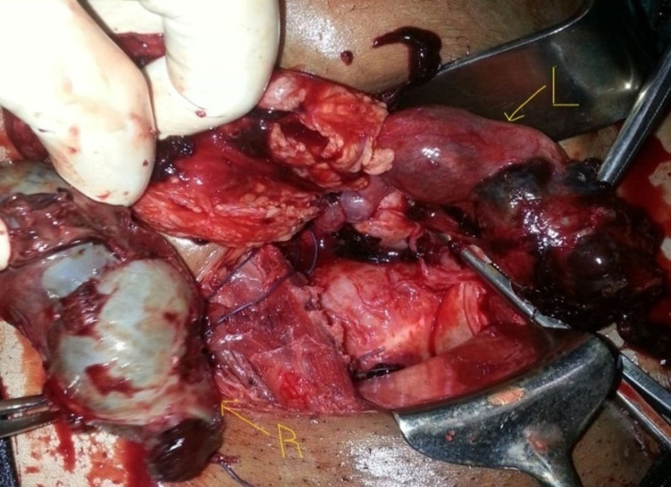
Bilateral tubal ectopic pregnancy is a very rare form of extra-uterine pregnancy with high maternal morbidity and mortality if intervention is delayed. We report the case of a 27-year-old para 2 gravida 3 patient who presented in haemorrhagic shock after delayed diagnosis of ectopic pregnancy. An ultrasound scan noted a right tubal ectopic pregnancy. At laparotomy, bilateral ruptured tubal ectopic pregnancy was encountered and bilateral salpingectomy was done as both tubes were not salvageable. She recovered completely postoperatively and histology confirmed bilateral tubal ectopic pregnancies. Bilateral tubal ectopic pregnancy may not be easily diagnosed on a scan; hence vigilance at surgery is critical to prevent maternal mortality.
Keywords: Spontaneous; bilateral; case report; ectopic pregnancy; ruptured.
Copyright: Michael Nyakura et al.
Conflict of interest statement
The authors declare no competing interests.
Figures


References
-
- Wakankar R, Kedar K. Ectopic pregnancy-a rising trend. International Journal of Scientific Study. 2015;3(5):18–19.
-
- Chinokwetu-Marere T. The prevalence and morbidity associated with ectopic pregnancies at Harare Central and Parirenyatwa Hospitals. University of Zimbabwe. 2013;3:18.
-
- Sommer EM, Reisenberger K, Bogner G, Nagele F. Laparoscopic management of an unrecognized spontaneous bilateral tubal pregnancy. Acta Obstet Gynecol Scand. 2002 Apr;81(4):366–8. - PubMed
-
- Edelstein MC, Morgan MA. Bilateral simultaneous tubal pregnancy: case report and review of the literature. Obstet Gynecol Surv. 1989;44(4):250–2. - PubMed
Publication types
MeSH terms
LinkOut - more resources
Full Text Sources
