Case Reports
. 2021 Jul 30:2021:9944886.
doi: 10.1155/2021/9944886. eCollection 2021.

Rare Anal Canal Cancer with Secondary Extramammary Paget's Disease (Pagetoid Spread) Complicated by Squamous Cell Carcinoma of the Skin

Affiliations
Case Reports

Rare Anal Canal Cancer with Secondary Extramammary Paget's Disease (Pagetoid Spread) Complicated by Squamous Cell Carcinoma of the Skin

Tatsuki Kusuhara et al. Case Rep Surg. .

Abstract

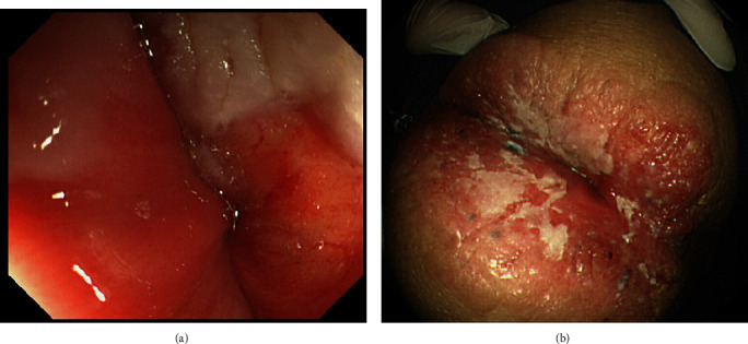
A 91-year-old man had a node and erythema in the anal area resistant to treatment. A biopsy of the node in the anus showed atypical cells developing as Paget's disease, and staining revealed that the cells were CK7-positive, CK20-positive, and GCDFP15-negative. Therefore, tumor invasion with pagetoid spread (PS) from the anus to the skin was suspected, and the patient was referred to our department for a close examination and surgical treatment. Lower gastrointestinal endoscopy showed edematous, hemorrhagic mucosa in the anal canal, and he was diagnosed with adenocarcinoma via a biopsy. Additionally, redness and swelling with white moss were observed on the skin around the anus. Biopsy showed that Paget cells were diffusely present in the epithelium, and an image of squamous cell carcinoma directly under the epithelium was obtained. Taken together, the patient was diagnosed with the invasion of anal canal cancer with PS to the skin, and we performed laparoscopic abdominoperineal resection and skin carcinoma resection in the perineum. The histopathological analysis showed adenocarcinoma invading the external anal sphincter and subcutaneous adipose tissue in the vicinity of the pectinate line of the anal canal. Pagetoid spread of the adenocarcinoma was observed in the epidermis, and the open portion was slightly invaded up to the rectal mucosa. The anal skin region of the adenocarcinoma partially continued to the hair follicles, and it was complicated by squamous cell carcinoma invading the dermis. There are a few reports of anal canal cancer with PS, and the coexistence of adenocarcinoma and squamous cell carcinoma, as seen in the present case, is rare. We report our case together with relevant literature.


Conflict of interest statement

The authors declare that there is no conflict of interest regarding the publication of this article.

Figures

Figure 1
Figure 1
Contrast-enhanced abdominal CT scan. Sagittal section. Enhanced abdominal CT shows a contrast effect on the perianal skin in the early phase.
Figure 2
Figure 2
Endoscopic photograph of the lower gastrointestinal tract. (a) The mucous membrane of the anal canal was edematous and bled easily. (b) Redness and swelling with white moss were observed in the anus.
Figure 3
Figure 3
Macroscopic image of the resected specimen surrounding the anus. There was a 92 mm × 48 mm tumor accompanied by slightly protruding white moss in the entire circumference of the anus with a clear boundary between the tumor and the surrounding healthy epithelium.
Figure 4
Figure 4
Histological images. (a) Histological image of the vicinity of the pectinate line of the anal canal showing the invasion of adenocarcinoma with tube formation directly under the stratified squamous epithelium. It was mucinous. Pagetoid cells, which are large clear cells, diffused into the stratified squamous epithelium. (b) Histological image of the area between the pectinate line and the opening on the rectal mucous side. Pagetoid spread of atypical cells was observed in the rectal epithelium. (c) A histological image of the squamous cell carcinoma around the anus. The anal skin region partially continued to the hair follicles, and it was complicated with well-differentiated squamous cell carcinoma invading the dermis.
Figure 5
Figure 5
Immunohistochemical analysis of the resected adenocarcinoma around the anus. (a) Immunostaining for CK7. The adenocarcinoma directly under the squamous epithelium and atypical cells developing in the squamous epithelium was positive for CK7. (b) Immunostaining for CK19. The adenocarcinoma directly under the squamous epithelium and atypical cells developing in the squamous epithelium was positive for CK19. (c) Immunostaining for CK20. The adenocarcinoma directly under the squamous epithelium and atypical cells developing in the squamous epithelium was positive for CK20. (d) Immunostaining for CDX2. The adenocarcinoma directly under the squamous epithelium and atypical cells developing in the squamous epithelium was positive for CDX2. (e) Immunostaining of GCDFP15. The adenocarcinoma directly under the squamous epithelium and atypical cells developing in the squamous epithelium was negative for GCDFP15. (f) Immunostaining for MUC5A. The adenocarcinoma directly under the squamous epithelium was partially positive for MUC5A.
Figure 6
Figure 6
Immunohistochemical analysis of the squamous cell carcinoma in the anal skin region. (a) Immunostaining for p63. The atypical squamous epithelium was positive for p63 in all layers. Pagetoid cells diffusely present in the epithelium were negative. (b) Immunostaining for CK5/6. The atypical squamous epithelium was positive for CK5/6 in all layers. Pagetoid cells diffusely present in the epithelium were negative.

Similar articles

Cited by

References

    1. Chanda J. J. Extramammary Paget’s disease: prognosis and relationship to internal malignancy. The American Journal of Dermatopathology. 1985;13(6):1009–1014. doi: 10.1016/S0190-9622(85)70254-X. - DOI - PubMed
    1. Claire K. S., Hoover A., Ashack K., Khachemoune A. Extramammary Paget disease. Dermatology Online Journal. 2019;25(4) doi: 10.5070/D3254043591. - DOI - PubMed
    1. Kitano A., Izumi M., Tamura K., Tonegawa M., Etou T., Kishida Y. Brain metastasis from cutaneous squamous cell carcinoma coexistent with extramammary Paget’s disease: a case report. Pathology International. 2019;69(10):619–625. doi: 10.1111/pin.12846. - DOI - PubMed
    1. Tanabe H., Kishigawa T., Sayama S., Tanaka T. A case of giant extramammary Paget’s disease of the genital area with squamous-cell carcinoma. Dermatology. 2001;202(3):249–251. doi: 10.1159/000051646. - DOI - PubMed
    1. Paget J. On disease of the mammary areola preceding cancer of the mammary gland. St. Bartholowmew. Hospital Reports. 1874;10:87–89.

Publication types