

Epiretinal membrane-induced intraretinal neovascularization
- PMID: 34381926
- PMCID: PMC8332659
- DOI: 10.1016/j.ajoc.2021.101180
Epiretinal membrane-induced intraretinal neovascularization
Abstract
Purpose: To report a 71-year-old male patient diagnosed with epiretinal membrane-induced intraretinal neovascularization.
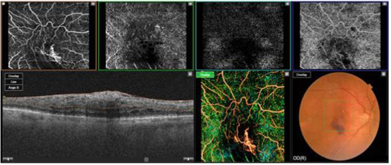
Observations: The presence of an epiretinal membrane (ERM) was confirmed by Optical Coherence Tomography (OCT), fluorescein and indocyanine angiography. Optical coherence tomography angiography (OCT-A) revealed a neovascular membrane within the ERM. Intravitreal ranibizumab injections were administered three times at four-week intervals. Imaging revealed a stable membrane with no leakage. Five months after the third injection, OCT revealed intraretinal fluid. OCT-A showed a new branch of the neo-vascular membrane at the superficial capillary plexus. Following an additional ranibizumab injection, the membrane stabilized.
Conclusions and importance: It is conceivable that neovascularization developed due to, or in close conjunction with an epiretinal membranes already in place.
Keywords: Epiretinal membrane; Intraretinal neovascularization; Optical coherence tomography; Optical coherence tomography angiography.
© 2021 The Authors. Published by Elsevier Inc.
Conflict of interest statement
The authors have no financial disclosures.
Figures


References
-
- Pearlstone A.D. The incidence of idiopathic preretinal macular gliosis. Ann Ophthalmol. 1985;17:378–380. - PubMed
-
- Schmitz-Valckenberg S., Holz F.G., Bird A.C., Spaide A.F. Fundus autofluorescence imaging: Review and perspectives. Retina. 2008;28:385–409. - PubMed
-
- Kadonosono K., Itoh N., Nomura E., Ohno S. Capillary blood flow velocity in patients with idiopathic epiretinal membranes. Retina. 1999;19:536–539. - PubMed
-
- Wise G.N. Clinical features of idiopathic preretinal macular fibrosis. Schoenberg Lecture. Am J Ophthalmol. 1975;79:349–357. - PubMed
-
- Coscas G.J., Lupidi M., Coscas F., Cagini C., Souied E.H. Optical coherence tomography angiography versus traditional multimodal imaging in accessing the activity of exudative age-related macular degenaration: a new diagnostic challenge. Retina. 2015;35:2219–2228. - PubMed
Publication types
LinkOut - more resources
Full Text Sources
