

Aptamer-mediated synthesis of multifunctional nano-hydroxyapatite for active tumour bioimaging and treatment
- PMID: 34382270
- PMCID: PMC8450118
- DOI: 10.1111/cpr.13105
Aptamer-mediated synthesis of multifunctional nano-hydroxyapatite for active tumour bioimaging and treatment
Abstract
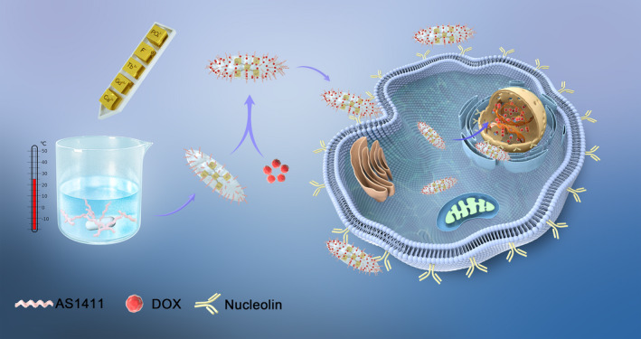
Objectives: The nano-hydroxyapatite (nHAp) is widely used to develop imaging probes and drug carriers due to its excellent bioactivity and biocompatibility. However, traditional methods usually need cumbersome and stringent conditions such as high temperature and post-modification to prepare the functionalized nHAp, which do not benefit the particles to enter cells due to the increased particle size. Herein, a biomimetic synthesis strategy was explored to achieve the AS1411-targeted tumour dual-model bioimaging using DNA aptamer AS1411 as a template. Then, the imaging properties and the biocompatibility of the synthesized AS-nFAp:Gd/Tb were further investigated.
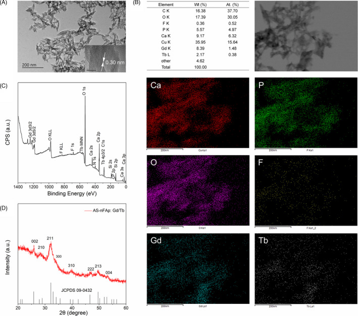
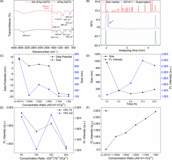
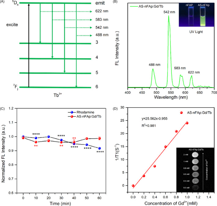
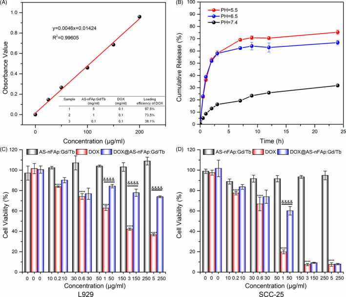
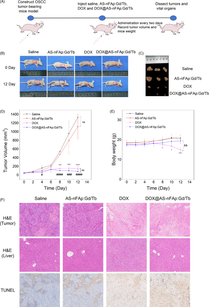
Materials and methods: The AS-nFAp:Gd/Tb was prepared under mild conditions through a one-pot procedure with AS1411 as a template. Besides, the anticancer drug DOX was loaded to AS-nFAp:Gd/Tb so as to achieve the establishment of a multifunctional nano-probe that integrated the tumour diagnosis and treatment. The AS-nFAp:Gd/Tb was characterized by transmission electron microscopy (TEM), energy disperse X-ray Spectroscopy (EDS) mapping, X-ray photoelectron spectroscopy (XPS) spectrum, X-ray diffraction (XRD), fourier-transformed infrared (FTIR) spectroscopy, capillary electrophoresis analyses, zeta potential and particle sizes. The in vitro magnetic resonance imaging (MRI) and fluorescence imaging were performed on an MRI system and a confocal laser scanning microscope, respectively. The potential of the prepared multifunctional nHAp for a targeted tumour therapy was investigated by a CCK-8 kit. And the animal experiments were conducted on the basis of the guidelines approved by the Animal Care and Use Committee of Sichuan University, China.
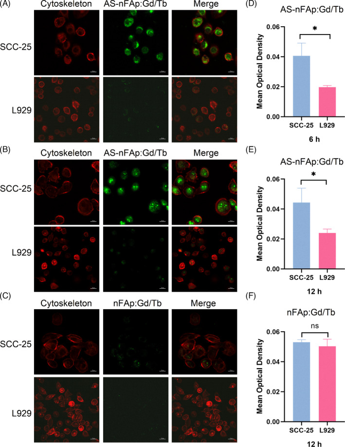
Results: In the presence of AS1411, the as-prepared AS-nFAp:Gd/Tb presented a needle-like morphology with good monodispersity and improved imaging performance. Furthermore, due to the specific binding between AS1411 and nucleolin up-expressed in cancer cells, the AS-nFAp:Gd/Tb possessed excellent AS1411-targeted fluorescence and MRI imaging properties. Moreover, after loading chemotherapy drug DOX, in vitro and in vivo studies showed that DOX@AS-nFAp:Gd/Tb could effectively deliver DOX to tumour tissues and exert a highly effective tumour inhibition without systemic toxicity compared with pure DOX.
Conclusions: The results indicated that the prepared multifunctional nHAp synthesized by a novel biomimetic strategy had outstanding capabilities of recognition and treatment for the tumour and had good biocompatibility; hence, it might have a potential clinical application in the future.
Keywords: AS1411; biomimetic synthesis; drug carriers; dual-model bioimaging; nano-hydroxyapatite.
© 2021 The Authors. Cell Proliferation published by John Wiley & Sons Ltd.
Conflict of interest statement
No conflict of interest was declared in this article.
Figures
References
-
- Zhen X, Zhang J, Huang J, Xie C, Miao Q, Pu K. Macrotheranostic probe with disease‐activated near‐infrared fluorescence, photoacoustic, and photothermal signals for imaging‐guided therapy. Angew Chem Int Ed Engl. 2018;57:7804‐7808. - PubMed
-
- Ma X, Zhao Y, Liang XJ. Theranostic nanoparticles engineered for clinic and pharmaceutics. Acc Chem Res. 2011;44:1114‐1122. - PubMed
-
- Abeylath SC, Ganta S, Iyer AK, Amiji M. Combinatorial‐designed multifunctional polymeric nanosystems for tumor‐targeted therapeutic delivery. Acc Chem Res. 2011;44:1009‐1017. - PubMed
-
- Gao S, Li Y, Xiao D, Zhou M, Cai X, Lin Y. Tetrahedral framework nucleic acids induce immune tolerance and prevent the onset of type 1 diabetes. Nano Lett. 2021;21:4437‐4446. - PubMed
-
- Liu Y, Sun Y, Li S, et al. Tetrahedral framework nucleic acids deliver antimicrobial peptides with improved effects and less susceptibility to bacterial degradation. Nano Lett. 2020;20:3602‐3610. - PubMed
MeSH terms
Substances
Grants and funding
LinkOut - more resources
Full Text Sources
Other Literature Sources
Medical
Research Materials
