. 2021 Oct;8(19):e2100584.
doi: 10.1002/advs.202100584. Epub 2021 Aug 11.

N2-Polarized Neutrophils Guide Bone Mesenchymal Stem Cell Recruitment and Initiate Bone Regeneration: A Missing Piece of the Bone Regeneration Puzzle

Affiliations

N2-Polarized Neutrophils Guide Bone Mesenchymal Stem Cell Recruitment and Initiate Bone Regeneration: A Missing Piece of the Bone Regeneration Puzzle

Bolei Cai et al. Adv Sci (Weinh). 2021 Oct.

Abstract

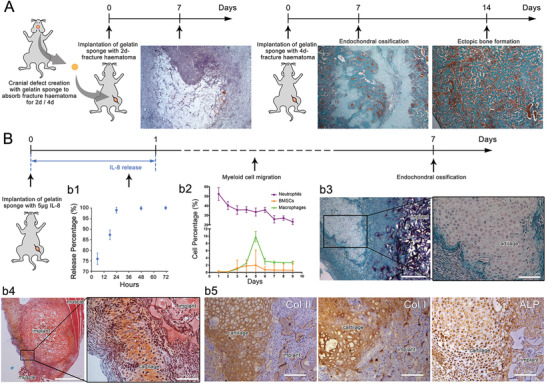
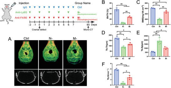
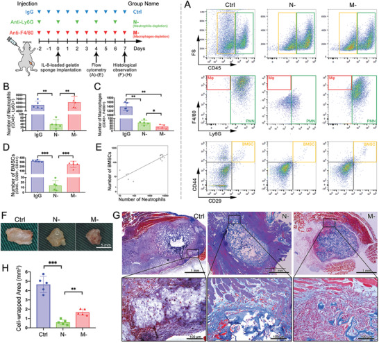
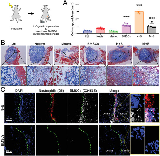
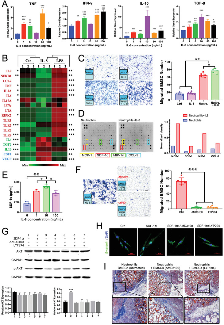
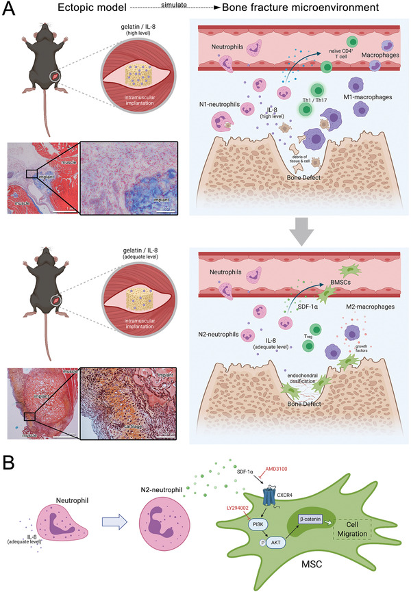
The role of neutrophils in bone regeneration remains elusive. In this study, it is shown that intramuscular implantation of interleukin-8 (IL-8) (commonly recognized as a chemotactic cytokine for neutrophils) at different levels lead to outcomes resembling those of fracture hematoma at various stages. Ectopic endochondral ossification is induced by certain levels of IL-8, during which neutrophils are recruited to the implanted site and are N2-polarized, which then secrete stromal cell-derived factor-1α (SDF-1α) for bone mesenchymal stem cell (BMSC) chemotaxis via the SDF-1/CXCR4 (C-X-C motif chemokine receptor 4) axis and its downstream phosphatidylinositol 3'-kinase (PI3K)/Akt pathway and β-catenin-mediated migration. Neutrophils are pivotal for recruiting and orchestrating innate and adaptive immunocytes, as well as BMSCs at the initial stage of bone healing and regeneration. The results in this study delineate the mechanism of neutrophil-initiated bone regeneration and interaction between neutrophils and BMSCs, and innate and adaptive immunities. This work lays the foundation for research in the fields of bone regenerative therapy and biomaterial development, and might inspire further research into novel therapeutic options.

Keywords: bone regeneration; interleukin-8; neutrophils; stem cell recruitment; stromal cell-derived factor-1α.


Conflict of interest statement

The authors declare no conflict of interest.

Figures

Figure 1
Figure 1
Intramuscular ectopic endochondral ossification induced by fracture hematoma and IL‐8 were similar at the early stage, recruiting neutrophils first and BMSCs and macrophages at a later stage. A) Histological observation of intramuscular implantation of 2‐day and 4‐day cranial defect hematoma using Masson's trichrome staining. 4‐day hematoma‐induced ectopic endochondral ossification. B) b1) In vitro release profile of IL‐8 from gelatin sponge. IL‐8 loaded onto gelatin sponge was rapidly released within 24 h. b2) Time‐course cytometric quantification of myeloid cell subpopulations in the implant using flow cytometry. Neutrophils promptly appeared and peaked at day 1, while BMSCs and macrophages peaked at day 5. Histological observation at day 7 using b3) Masson's trichrome staining and b4) Safranin‐O staining. Ectopic cartilage was formed around the implant. b5) Type II collagen (Col II), type I collagen (Col I), and alkaline phosphatase (ALP) immunohistochemistry staining. The exclusive cartilaginous Col II, together with the osteogenic‐related Col I and ALP indicated ectopic endochondral ossification. (n = 3 for each group)
Figure 2
Figure 2
Neutrophils were pivotal for bone‐healing at the initiating stage. α‐Ly6G or α‐F4/80 antibody was continuously injected from 2 days pre‐ to 7 days post‐cranial defect creation for depletion of neutrophils or macrophages (referred to as N‐ and M‐), respectively, with IgG antibody as a control. A) Micro‐CT reconstruction of the defect site after 90 days. Quantification histogram of B) bone volume/total volume (BV/TV), C) bone mineral density (BMD), D) trabecular thickness (Tb.Th.), E) trabecular space (Tb.Sp.), and F) trabecular number (Tb.N.). (** p < 0.01, * p < 0.05; n = 3 for each group)
Figure 3
Figure 3
Depletion of circulatory neutrophils cut off the IL‐8‐induced myeloid cell recruitment and ectopic endochondral ossification. A) Flow cytometry of the ectopic implant and its quantitative histogram of B) neutrophils, C) macrophages, and D) BMSCs. Depletion of neutrophils inhibited recruitment of all myeloid cells. E) Numbers of neutrophils and BMSCs exhibited a positive correlation. F) Implants retrieved at day 7, G) histological observation using Masson's trichrome staining, and H) quantification of cell‐enwrapped areas. Control group induced ectopic cartilage formation, while N‐ and M‐ significantly decreased the cell‐wrapped area and prevented further differentiation. (*** p < 0.001, ** p < 0.01, * p < 0.05; n = 5 for each group)
Figure 4
Figure 4
Interaction of neutrophils and BMSCs were key to anabolism, while macrophages were essential for catabolism. Irradiated mice received IL‐8‐gelatin implantation and injection of exogenous myeloid cells. A) Quantification of cell‐enwrapped areas and B) histological observation using Masson's trichrome staining. C) Fluorescent‐labeled neutrophils and BMSCs around the implant. When neutrophils and BMSCs were simultaneously transfused (N+B), greater cell recruitment and ECM were observed around the implant; when only macrophages were transfused the implant was rapidly degraded. (Compared with ctrl: *** p < 0.001, ** p < 0.01, * p < 0.05; n = 5 for each group).
Figure 5
Figure 5
IL‐8 (10 ng mL−1) polarized neutrophils toward the N2 phenotype, which secreted SDF‐1α to mediate BMSC recruitment and differentiation via the SDF‐1/CXCR4 axis and its downstream PI3K/AKT pathway and β‐catenin‐mediated migration. A) Expressions of proinflammatory and anti‐inflammatory genes in neutrophils under different concentrations of IL‐8. B) Heat map of mouse inflammatory response and autoimmunity profiler PCR array: Untreated (Ctr), IL‐8‐, and LPS‐treated neutrophils (10 ng mL−1, 24 h exposure). Data are row‐relative and normalized to GAPDH. C) Transwell assay highlighting BMSC recruitment by IL‐8, neutrophils, and IL‐8‐treated neutrophils (migrated cells counted in five random 200 × microscopic fields). D) Mouse cytokine antibody array analyses of conditioned media from neutrophils or IL‐8‐treated neutrophils (10 ng mL−1, 24 h exposure) and the top four upregulated cytokines quantified using densitometry. E) ELISA‐determined SDF‐1α secreted by neutrophils treated with IL‐8 at different concentrations. F) Recruitment of BMSC pretreated with CXCR4 antagonist AMD3100 and PI3K/AKT inhibitor LY294002. G) SDF‐1‐induced phosphorylation of Akt was inhibited by AMD3100 or LY294002. H) Immunofluorescent staining of β‐catenin translocated to nuclei by SDF‐1α and inhibited by AMD3100 or LY294002. I) Irradiated mice received IL‐8‐gelatin implantation and injection of exogenous neutrophils and BMSCs pretreated with AMD3100 or LY294002. (Compared with ctrl: *** p < 0.001, ** p < 0.01, * p < 0.05. Compared with 10 ng mL−1: ### p < 0.001, ## p < 0.01, # p < 0.05; n = 3 for each group in PCR and ELISA; n = 5 for each group in transwell assay).
Figure 6
Figure 6
A) The role of neutrophils in the initiation process of bone regeneration elucidated via in vivo ectopic simulation of endochondral ossification. The ectopic implantation of IL‐8 at a high and medium level have simulated the microenvironment of bone fracture at the early and later stage, respectively. High‐level IL‐8 at the early stage led to a proinflammatory microenvironment and phagocytosis to eliminate the damaged tissue debris for inflammation resolution. Thereafter, an adequate level of IL‐8 at the later stage polarizes neutrophils to an N2‐subtype for endochondral ossification. B) Signaling pathway of BMSC recruitment by neutrophils. N2‐polarized neutrophils release SDF‐1α to recruit BMSCs via the SDF‐1/CXCR4 axis, its downstream PI3K/Akt pathway, and β‐catenin‐mediated migration.

References

    1. Wells J. M., Watt F. M., Nature 2018, 557, 322. - PubMed
    1. a) Petite H., Viateau V., Bensaid W., Meunier A., de Pollak C., Bourguignon M., Oudina K., Sedel L., Guillemin G., Nat. Biotechnol. 2000, 18, 959; - PubMed
    2. b) Harada S., Rodan G. A., Nature 2003, 423, 349; - PubMed
    3. c) Grayson W. L., Bunnell B. A., Martin E., Frazier T., Hung B. P., Gimble J. M., Nat. Rev. Endocrinol. 2015, 11, 140. - PMC - PubMed
    1. Madl C. M., Heilshorn S. C., Blau H. M., Nature 2018, 557, 335. - PMC - PubMed
    1. a) Neves J., Zhu J., Sousa‐Victor P., Konjikusic M., Riley R., Chew S., Qi Y., Jasper H., Lamba D. A., Science 2016, 353, aaf3646; - PMC - PubMed
    2. b) Buckley C. D., Gilroy D. W., Serhan C. N., Stockinger B., Tak P. P., Nat. Rev. Immunol. 2013, 13, 59. - PubMed
    1. Forbes S. J., Rosenthal N., Nat. Med. 2014, 20, 857. - PubMed

Publication types

LinkOut - more resources