. 2021 Sep;20(17):1629-1641.
doi: 10.1080/15384101.2021.1924450. Epub 2021 Aug 12.

Role of lncRNA XIST/microRNA-19/PTEN network in autophagy of nucleus pulposus cells in intervertebral disc degeneration via the PI3K/Akt signaling pathway

Affiliations

Role of lncRNA XIST/microRNA-19/PTEN network in autophagy of nucleus pulposus cells in intervertebral disc degeneration via the PI3K/Akt signaling pathway

Wei Chen et al. Cell Cycle. 2021 Sep.

Abstract

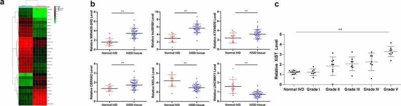
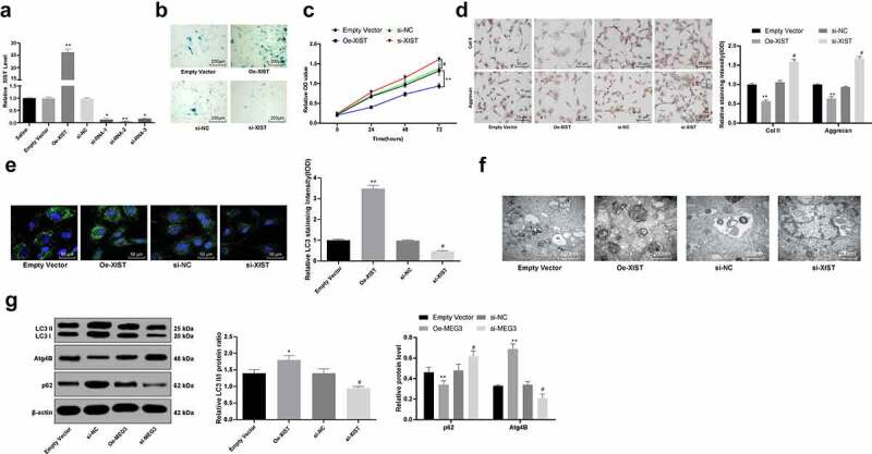
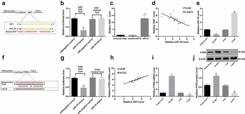
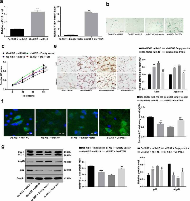
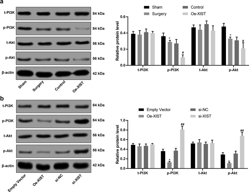
Intervertebral disc degeneration (IVDD) is a complicated pathological condition accompanying with low back pain. This study was designed to figure out the mechanism of lncRNA XIST in IVDD. Abnormally expressed lncRNAs in IVDD patients were measured. The correlations among XIST, miR-19 and PTEN were identified. Overexpression and silencing of XIST, miR-19 and PTEN were introduced and their roles in NPC autophagy in vitro were detected. The potential signaling pathway involved in these events was identified. Consequently, high expression of XIST was found in IVDD patients. It induced NPC autophagy and reduced NPC viability. XIST could serve as a competing endogenous RNA (ceRNA) for miR-19 and upregulate PTEN expression. The overexpression of XIST reduced miR-19 expression, which was followed by enhanced PTEN expression. Upregulation of miR-19 increased NPC viability and proliferation, while decreased NPC autophagy that regulated by XIST, while overexpressed PTEN reversed the above changes. Moreover, overexpression of XIST inactivated the PI3k/Akt signaling pathway.

Keywords: Intervertebral disc degeneration; PTEN; autophagy; competing endogenous RNA; long non-coding RNA XIST; microRNA-19; nucleus pulposus cells.


Conflict of interest statement

The authors declared that they have no competing interests.

Figures

Figure 1.
Figure 1.
LncRNA XIST is upregulated in IVDD patients in a Pfirrmann grade-dependent manner. (a), heatmap for differentially expressed lncRNAs between 10 IVD tissue samples from 5 IVDD patients and 5 non-IVDD patients using microarray analysis; (b), randomly selected 6 differentially expressed lncRNAs (MIR4435-2HG, lncRNA:iab8, ATXN8OS, CERNA2, NKILA and LINC00511) identified using RT-qPCR; (c), lncRNA XIST expression in 36 IVDD patients with different Pfirrmann grades (1–5) and 12 normal IVD controls (0) measured using RT-qPCR. Data were analyzed by the t test or one-way ANOVA, followed by Tukey’s multiple comparison test; * p < 0.05, compared to the IVD normal controls
Figure 2.
Figure 2.
Silencing XIST enhances NPC viability and inhibits autophagy in vitro. (a), XIST expression in NPCs measured using RT-qPCR after transfection; (b), senescence of each group of NPCs detected by SA-β-gal staining; (c), viability of NPCs assessed using MTT assay; (d), levels of Col II and aggrecan in NPCs assessed using immunocytochemistry; (e), LC3 expression in NPCs confirmed by immunofluorescence staining; (f), formation of autophagosomes in NPCs observed under a TEM; (g), protein levels of LC3II, LC3I, p62 and Atg4B evaluated using western blot analysis. Each experiment was performed three times independently. Data are expressed as the mean ± standard deviation (n = 3); Data were analyzed by one-way, followed by Tukey’s multiple comparison test; *, compared to the empty vector group, p < 0.05; # compared to the si-NC group, p < 0.05
Figure 3.
Figure 3.
XIST serves as a ceRNA for miR-19 to regulate PTEN expression. (a), binding relation between miR-19 and XIST predicted on a bio-information website (http://www.mircode.org/); (b,c), binding relation between miR-19 and XIST identified by dual luciferase reporter gene assay (b) and RNA-pull down assay (c); (d), plot analysis of the correlation between XIST and miR-19 in 36 IVDD patients; (e), relative expression of miR-19 determined by RT-qPCR; (f), the binding relation between miR-19 and PTEN predicted via TargetScan (http://www.targetscan.org/vert_72/); (g), binding relation between miR-19 and PTEN identified using dual luciferase reporter gene assay; (h), plot analysis of the correlation between miR-19 and PTEN in 36 IVDD patients; (i,j), relative mRNA expression (i) and protein level of PTEN (j) measured using RT-qPCR and western blot analysis, respectively; Each experiment was performed three times independently. Data were analyzed by one-way, followed by Tukey’s multiple comparison test. In panels (d) and (h), Pearson’s correlation coefficient test was utilized for statistical analysis, n = 45, *, compared to the empty vector group, p < 0.05, #, compared to the si-NC group, p < 0.05
Figure 4.
Figure 4.
Overexpression of miR-19 increases NPC viability and decreases cell autophagy regulated by XIST. (a), the expression of miR-19 and PTEN mRNA measured using RT-qPCR after transfection; (b), senescence of NPCs detected with SA‐β‐gal staining; (c), viability of NPCs assessed using MTT assay; (d), levels of Col II and aggrecan in NPCs assessed using immunocytochemistry; (e), LC3 expression in NPCs confirmed with immunofluorescence staining; (f), protein levels of LC3I, LC3II, p62 and Atg4B evaluated using western blot analysis. Data are expressed as the mean ± standard deviation. Each experiment was performed three times independently. Data were analyzed by the t test or one-way ANOVA, followed by Tukey’s multiple comparison test. *, compared to the empty vector group, p < 0.05, # compared to the si-NC group, p < 0.05; Repetition = 3
Figure 5.
Figure 5.
XIST inactivates the PI3K/Akt signaling pathway. (a), PI3K and Akt levels in IVD tissues measured using western blot analysis; (b), protein levels of PI3K and Akt in NPCs measured using western blot analysis; Each experiment was performed three times independently. The data are expressed as the mean ± standard deviation. Data were analyzed by one-way ANOVA, followed by Tukey’s multiple comparison test. *, compared to the sham or empty vector group, p < 0.05, # compared to the control group, p < 0.05, ## compared to the si-NC group, p < 0.01

References

    1. Zhang Z, Xu T, Chen J, et al. Parkin-mediated mitophagy as a potential therapeutic target for intervertebral disc degeneration. Cell Death Dis. 2018;9:980. - PMC - PubMed
    1. Li X, Lou Z, Liu J, et al. Upregulation of the long noncoding RNA lncPolE contributes to intervertebral disc degeneration by negatively regulating DNA polymerase epsilon. Am J Transl Res. 2019;11:2843–2854. - PMC - PubMed
    1. Sampara P, Banala RR, Vemuri SK, et al. Understanding the molecular biology of intervertebral disc degeneration and potential gene therapy strategies for regeneration: a review. Gene Ther. 2018;25:67–82. - PubMed
    1. Yang Q, Guo XP, Cheng YL, et al. MicroRNA-143-5p targeting eEF2 gene mediates intervertebral disc degeneration through the AMPK signaling pathway. Arthritis Res Ther. 2019;21:97. - PMC - PubMed
    1. Liu P, Chang F, Zhang T, et al. Downregulation of microRNA-125a is involved in intervertebral disc degeneration by targeting pro-apoptotic Bcl-2 antagonist killer 1. Iran J Basic Med Sci. 2017;20:1260–1267. - PMC - PubMed

MeSH terms