

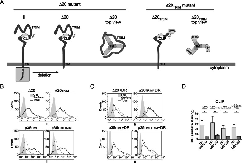
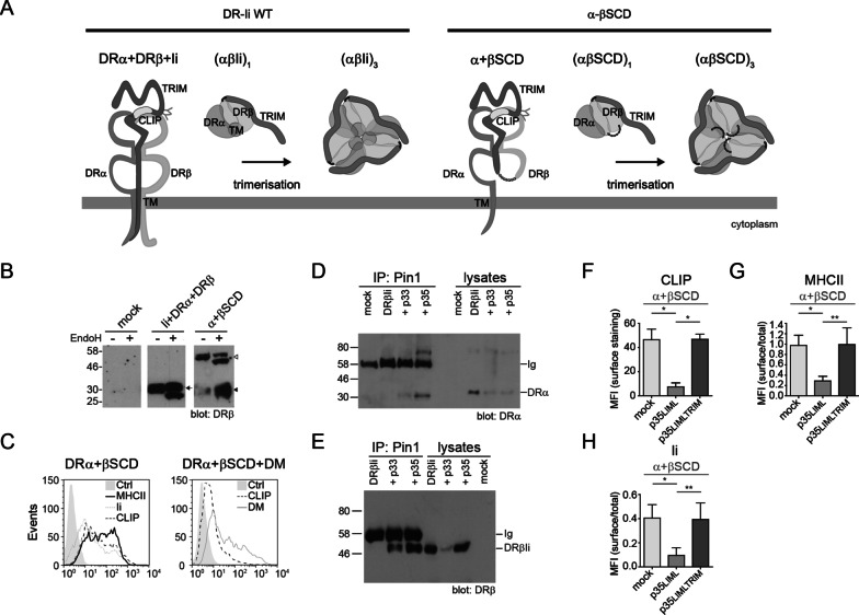
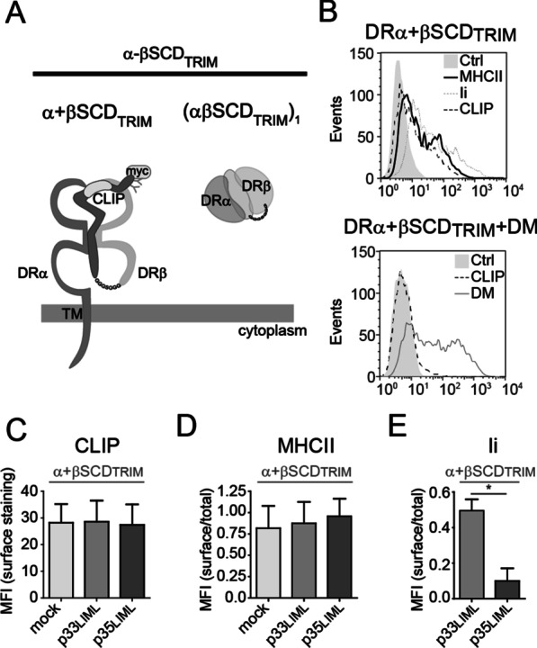
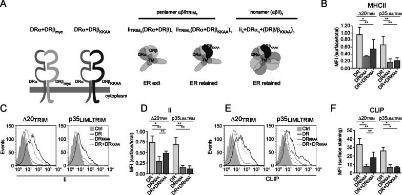
The transmembrane domain and luminal C-terminal region independently support invariant chain trimerization and assembly with MHCII into nonamers
- PMID: 34384367
- PMCID: PMC8362237
- DOI: 10.1186/s12865-021-00444-6
The transmembrane domain and luminal C-terminal region independently support invariant chain trimerization and assembly with MHCII into nonamers
Abstract
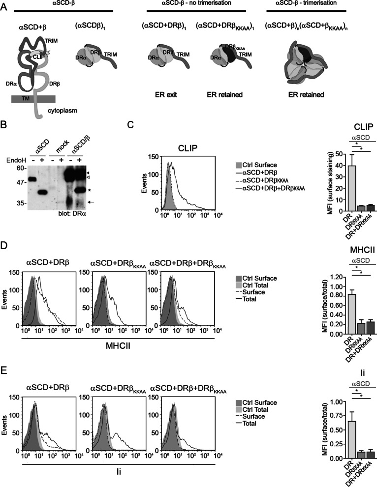
Background: Invariant chain (CD74, Ii) is a multifunctional protein expressed in antigen presenting cells. It assists the ER exit of various cargos and serves as a receptor for the macrophage migration inhibitory factor. The newly translated Ii chains trimerize, a structural feature that is not readily understood in the context of its MHCII chaperoning function. Two segments of Ii, the luminal C-terminal region (TRIM) and the transmembrane domain (TM), have been shown to participate in the trimerization process but their relative importance and impact on the assembly with MHCII molecules remains debated. Here, we addressed the requirement of these domains in the trimerization of human Ii as well as in the oligomerization with MHCII molecules. We used site-directed mutagenesis to generate series of Ii and DR mutants. These were transiently transfected in HEK293T cells to test their cell surface expression and analyse their interactions by co-immunoprecipitations.
Results: Our results showed that the TRIM domain is not essential for Ii trimerization nor for intracellular trafficking with MHCII molecules. We also gathered evidence that in the absence of TM, TRIM allows the formation of multi-subunit complexes with HLA-DR. Similarly, in the absence of TRIM, Ii can assemble into high-order structures with MHCII molecules.
Conclusions: Altogether, our data show that trimerization of Ii through either TM or TRIM sustains nonameric complex formation with MHCII molecules.
Keywords: Antigen presentation; CD74; MHCII; Nonamerization; RXR; Transmembrane domain; Trimerization domain.
© 2021. The Author(s).
Conflict of interest statement
The authors declare that they have no competing interests.
Figures

References
-
- Jones PP, Murphy DB, Hewgill D, McDevitt HO. Detection of a common polypeptide chain in I-A and I–E sub-region immunoprecipitates. Mol Immunol. 1979;16:51–60. - PubMed
Publication types
MeSH terms
Substances
LinkOut - more resources
Full Text Sources
Research Materials
