. 2021 Aug 12;19(1):243.
doi: 10.1186/s12951-021-00975-5.

Peptide vaccine-conjugated mesoporous carriers synergize with immunogenic cell death and PD-L1 blockade for amplified immunotherapy of metastatic spinal

Affiliations

Peptide vaccine-conjugated mesoporous carriers synergize with immunogenic cell death and PD-L1 blockade for amplified immunotherapy of metastatic spinal

Zhenqing Wang et al. J Nanobiotechnology. .

Abstract

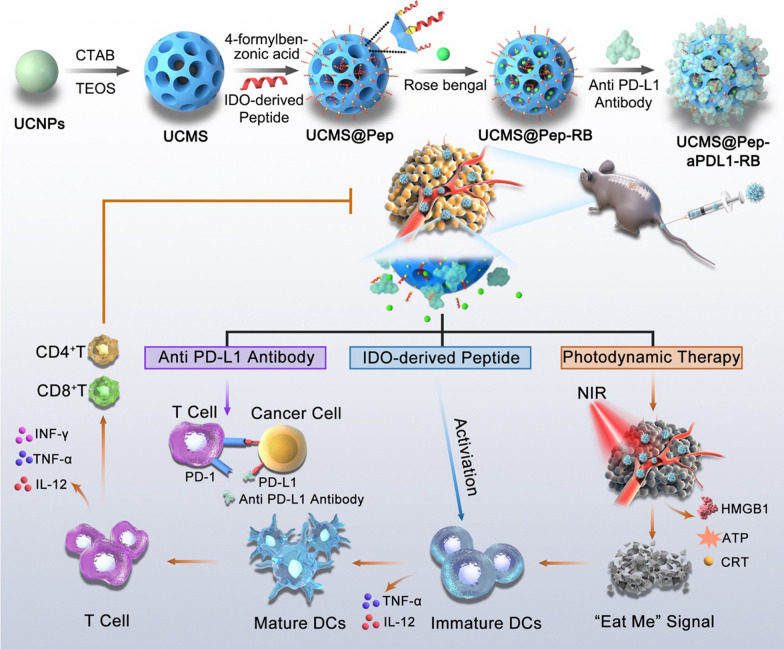
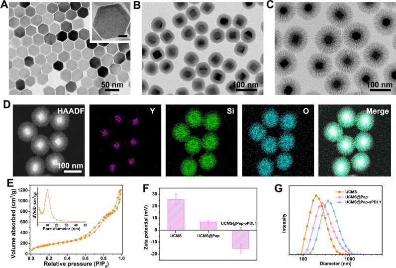
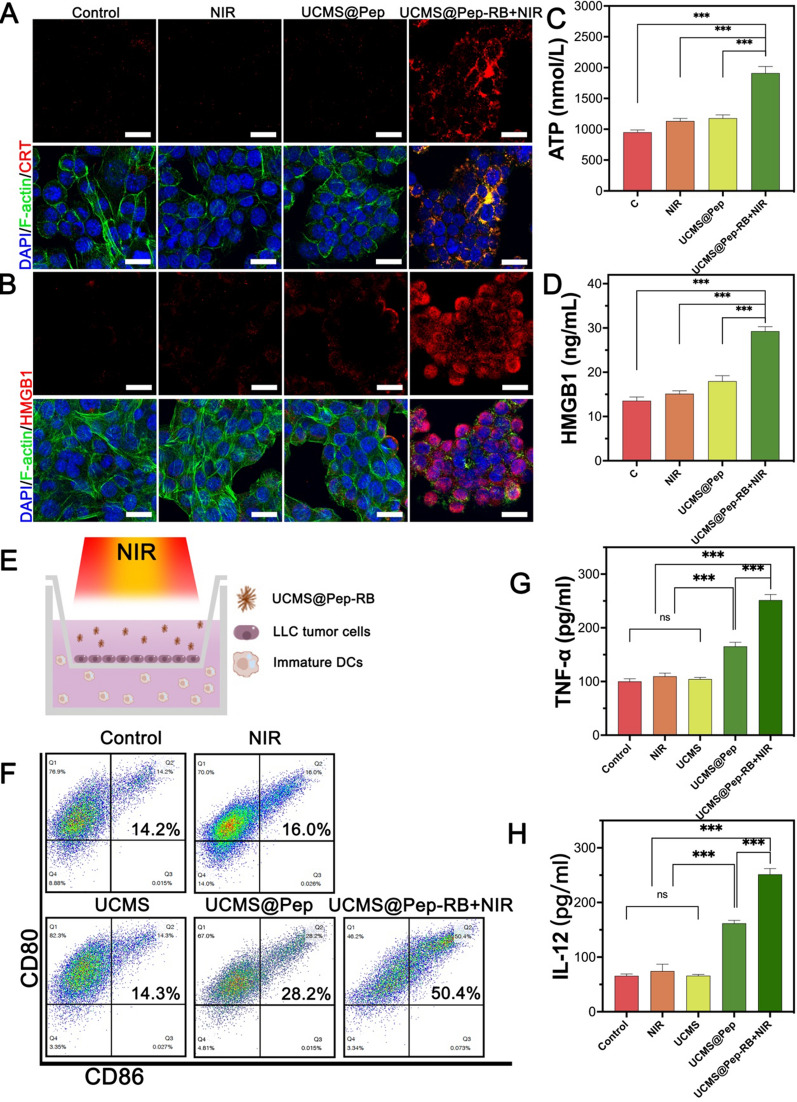
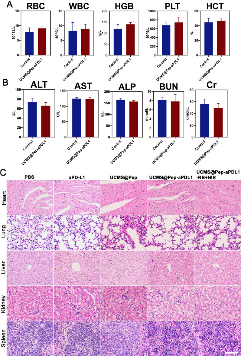
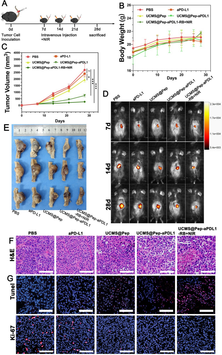
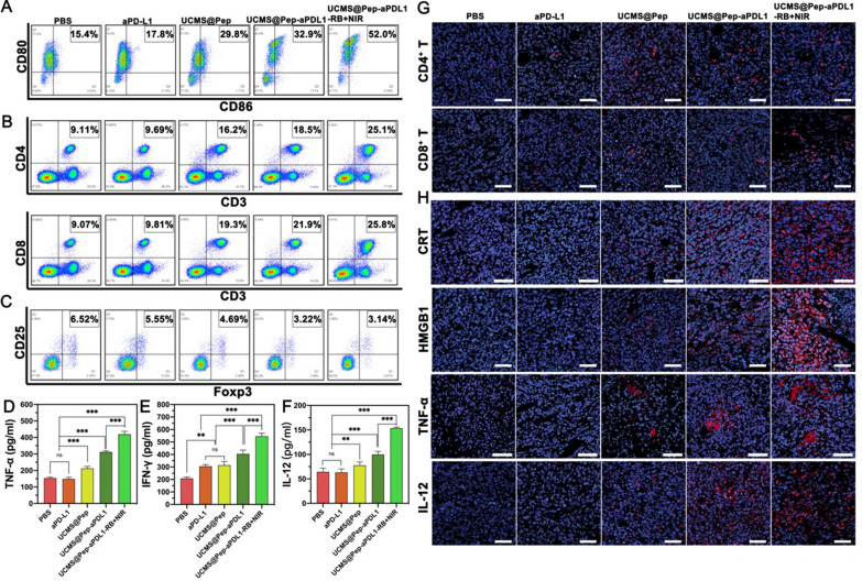
The clinical treatment of metastatic spinal tumor remains a huge challenge owing to the intrinsic limitations of the existing methods. Programmed cell death protein 1 (PD1)/programmed cell death ligand 1 (PD-L1) pathway blockade has been explored as a promising immunotherapeutic strategy; however, their inhibition has a low response rate, leading to the minimal cytotoxic T cell infiltration. To ameliorate the immunosuppressive microenvironment of intractable tumor and further boost the efficacy of immunotherapy, we report an all-round mesoporous nanocarrier composed of an upconverting nanoparticle core and a large-pore mesoporous silica shell (UCMS) that is simultaneously loaded with photosensitizer molecules, the IDO-derived peptide vaccine AL-9, and PD-L1 inhibitor. The IDO-derived peptide can be recognized by the dendritic cells and presented to CD8+ cytotoxic T cells, thereby enhancing the immune response and promoting the killing of the IDO-expressed tumor cells. Meanwhile, the near-infrared (NIR) activated photodynamic therapy (PDT) could induce immunogenic cell death (ICD), which promotes the effector T-cell infiltration. By combining the PDT-elicited ICD, peptide vaccine and immune checkpoint blockade, the designed UCMS@Pep-aPDL1 successfully potentiated local and systemic antitumor immunity and reduced the progression of metastatic foci, demonstrating a synergistic strategy for cancer immunotherapy.

Keywords: Immunogenic cell death; Peptide vaccine; Photodynamic therapy; Programmed cell death protein 1/programmed cell death ligand 1 (PD-1/PD-L1) blockades; Spine metastasis.


Conflict of interest statement

The authors declare that they have no known competing financial interests or personal relationships that could have appeared to influence the work reported in this paper.

Figures

Fig. 1
Fig. 1
Schematic illustration of the preparation of functional nanocarrier for photodynamic and peptide vaccine-amplified immunotherapy of spinal tumor
Fig. 2
Fig. 2
TEM images of (A) UCNPs (inset shows the corresponding high-magnification image; scale bar = 10 nm), B UCNPs@SiO2, and (C) UCMS. D High-angle annular dark field (HAADF) scanning TEM image and element mapping of the prepared UCMS. E Nitrogen adsorption isotherm of the prepared UCMS. F Zeta potential and (G) size distributions of UCMS, UCMS@Pep, and UCMS@Pep-aPDL1
Fig. 3
Fig. 3
In vitro photon conversion and cellular cytotoxicity of UCMS@Pep. A UV-vis spectrum of UCMS@Pep-RB (red line) and up-conversion emission spectrum of UCMS@Pep under excitation at 980 nm (green color). B Effects of irradiation time on the fluorescence intensity change of SOSG at 525 nm for different samples. C Relative viability of LLC cells incubated with different concentrations of blank nanoparticles, UCMS@Pep alone, and UCMS@Pep with NIR laser. D Results of calcein-AM & PI staining that was used to discriminate between live and dead cells (a: Control, b: NIR laser, c: UCMS@Pep, d: UCMS@Pep-RB + NIR laser, scale bar = 100 μm). (E Flow cytogram showing the results of the apoptosis assay based on the annexin V-FITC and propidium iodide (PI) staining of LLC cells after different treatments. F Corresponding inverted fluorescence microscopy images of DCFH-DA probe–stained LLC cells used to evaluate the overall intracellular ROS generation (scale bar = 50 μm). (*P < 0.05, **P < 0.01, ***P < 0.001, n = 3)
Fig. 4
Fig. 4
Intracellular ICD marker and DCs maturation of UCMS@Pep in vitro. A CRT exposure and (B) HMGB1 analysis by immunofluorescence using CLSM (scale bar = 25 μm). Release of (C) ATP and (D) HMGB1 detected by ELISA kits for different interventions. E Schematic of the trans-well DCs maturation system. F Flow cytometry results for mature DCs after different treatments. Levels of cytokines (G) TNF-α and (H) IL-12 in the culture medium. (*P < 0.05, **P < 0.01, ***P < 0.001, n = 3)
Fig. 5
Fig. 5
Biosafety in vivo of UCMS@Pep-aPDL1. A Results of routine blood count in mice treated with PBS and UCMS@Pep-aPDL1 (n ≥ 3); B Biochemistry indexes in mice treated with PBS and UCMS@Pep-aPDL1 (n ≥ 3); C H&E staining results obtained for the major organs of mice subjected to different treatments (Scale bar = 100 μm)
Fig. 6
Fig. 6
In vivo amplified anti-tumor effect of UCMS@Pep-aPDL1. A Schematics of the program used for spine metastasis tumor mode and IDO-based peptide tumor vaccine-mediated synergetic cancer treatment. Time-dependent (B) body weight and (C) tumor volume of mice in different groups. D Tumor progression in different groups evaluated by IVIS. E Tumor specimens extracted after the mice had been sacrificed. F H&E-stained histological images, G TUNEL-stained pathological changes and Ki-67-stained cellular proliferation in tumor tissues (scale bars = 100 μm). (*P < 0.05, **P < 0.01, ***P < 0.001, n > 3)
Fig. 7
Fig. 7
In vivo immune system enhancement by UCMS@Pep-aPDL1. AC Flow cytometric analyses of DCs, CD4 and CD8 T cells and Treg cells in the tumor tissues of mice immunized using different interventions. Secretion levels of (D) TNF-α, E IFN-γ, and F IL-12 in the serum of peripheral blood from treated mice. G Infiltration of CD4 and CD8 T cells in tumor sites detected by CLSM (scale bar = 50 μm). H Expression of CRT, HMGB1, and the infiltration of cytokines TNF-α and IL-12 in tumor sites evaluated by CLSM (scale bar = 50 μm). (*P < 0.05, **P < 0.01, ***P < 0.001, n > 3)

Similar articles

Cited by

References

    1. Siegel RL, Miller KD, Jemal A. Cancer statistics, 2020. CA Cancer J Clin. 2020;70(1):7–30. doi: 10.3322/caac.21590. - DOI - PubMed
    1. Amelot A, et al. Spinal metastases from lung cancer: survival depends only on genotype, neurological and personal status, scarcely of surgical resection. Surg Oncol. 2020;34:51–6. doi: 10.1016/j.suronc.2020.03.005. - DOI - PubMed
    1. Kuchuk M, et al. The incidence and clinical impact of bone metastases in non-small cell lung cancer. Lung Cancer. 2015;89(2):197–202. doi: 10.1016/j.lungcan.2015.04.007. - DOI - PubMed
    1. Silva GT, Bergmann A, Thuler LC. Incidence, associated factors, and survival in metastatic spinal cord compression secondary to lung cancer. Spine J. 2015;15(6):1263–9. doi: 10.1016/j.spinee.2015.02.015. - DOI - PubMed
    1. Sugiura H, et al. Predictors of survival in patients with bone metastasis of lung cancer. Clin Orthop Relat Res. 2008;466(3):729–36. doi: 10.1007/s11999-007-0051-0. - DOI - PMC - PubMed

MeSH terms