. 2021 Aug 12;14(1):39.
doi: 10.1186/s13072-021-00413-8.

Histone deacetylase inhibition leads to regulatory histone mark alterations and impairs meiosis in oocytes

Affiliations

Histone deacetylase inhibition leads to regulatory histone mark alterations and impairs meiosis in oocytes

Louis Legoff et al. Epigenetics Chromatin. .

Abstract

Background: Panobinostat (PB), a histone deacetylase (HDAC) inhibitor drug, is clinically used in the treatment of cancers. We investigated the effects of PB on murine ovarian functions in embryos and adult animals.

Methods: C57BL/6J mice were treated with 5 mg/kg PB on alternate days from embryonic day (E) 6.5 to E15.5. We analysed the effects of PB on the ovaries by using immunofluorescence, gene expression analysis and DNA methylation analysis techniques.

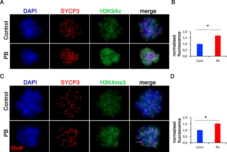
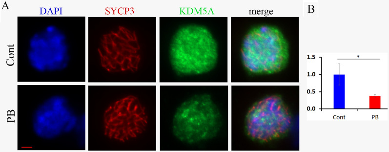
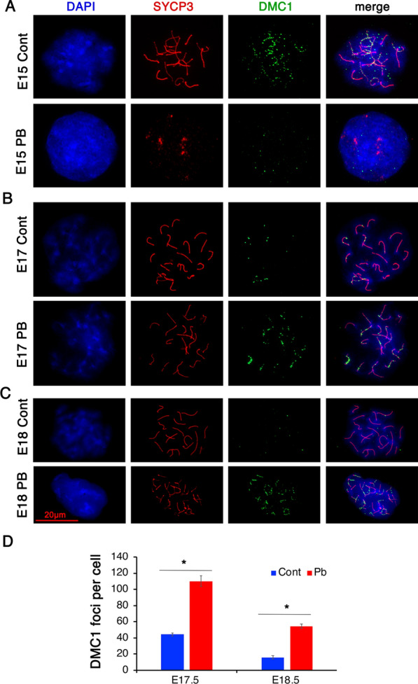
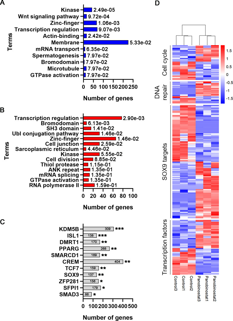
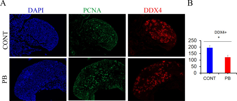
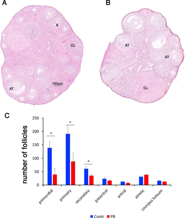
Results: At E15.5, we observed increases in histone H3K9Ac, H4Ac and H3K4me3 marks, while the level of the silencing H3K9me3 mark decreased. Synaptonemal complex examination at E15.5, E17.5 and E18.5 showed a delay in meiotic progression characterized by the absence of synaptonemal complexes at E15.5 and the persistence of double-strand breaks (DSBs) at E17.5 and E18.5 in PB-exposed oocytes. We found that exposure to PB led to changes in the expression of 1169 transcripts at E15.5. Genes regulated by the male-specific factors SRY-Box Transcription Factor 9 (SOX9) and Doublesex and Mab-3-related Transcription factor 1 (DMRT1) were among the most upregulated genes in the ovaries of PB-exposed mice. In contrast, PB treatment led to decreases in the expression of genes regulated by the WNT4 pathway. Notably, we observed 119 deregulated genes encoding Zn-finger proteins. The observed alterations in epigenetic marks and gene expression correlated with decreases in the numbers of germ cells at E15.5. After birth, PB-exposed ovaries showed increased proliferation of primary and secondary follicles. We also observed decreases in the numbers of primordial, primary and secondary follicles in adult ovaries from mice that were exposed to PB in utero. Finally, epigenetic alterations such as decreased H3K4me3 and increased H4 acetylation levels were also detected in somatic cells surrounding fully grown oocytes.

Conclusion: Our data suggest that inhibition of histone deacetylase by PB during a critical developmental window affects reprogramming and germ cell specification via alteration of epigenetic marks.

Keywords: Epigenetics; HDAC; Histone modifications; Ovary; Panobinostat; RNA-seq.


Conflict of interest statement

The authors declare that they have no competing interests.

Figures

Fig. 1
Fig. 1
Meiosis defects are associated with increases in histone acetylation and H3K4me3. A Surface spreads from E18.5 ovaries from control (first row) and Pb-exposed mice (second row) were immunostained with anti-H3K9Ac (green) and anti-SYCP3 (red) antibodies (63X magnification). The dotted circles represent telomere connections. B Quantitative analysis of immunofluorescence intensity. The data are presented as the averaged fluorescence compared to the control ± SEM; n = 4 control, n = 4 PB group; *p < 0.05, nonparametric Mann–Whitney test. C Surface spreads from E185 ovaries from control (first row) and PB-exposed mice (second row) were immunostained with anti-H3K4me3 (green) and anti-SYCP3 (red) antibodies; the arrows represent telomere end-to-end connections. D Quantitative analysis of anti-H3K4me3 intensities. The analysis was performed on at least 20 oocytes from four replicates for each group; n = 4 for each group; *p < 0.05, nonparametric Mann–Whitney test
Fig. 2
Fig. 2
KDM5A is decreased in exposed oocytes. Structurally preserved nuclei from E18,5 ovaries from the control (top row) and PB-exposed (bottom row) groups were immunostained for KDM5A and SYCP3. The combined total corrected fluorescence for KDM5A was calculated for each cell and normalized to the DAPI signal. The average normalized CTCF values are presented in MS-Excel plot ± SD; *p < 0.05, nonparametric Mann–Whitney test. The bar represents 2 μm
Fig. 3
Fig. 3
A critical meiotic step is perturbed in embryonic ovaries exposed to PB. Surface spreads from E15.5 control (A, top row) and PB-treated ovaries (A, bottom row) were immunostained with anti-DMC1 (green) and anti-SYCP3 (red) antibodies (63X magnification). B Surface spreads from E17.5 control (B, top row) and PB-treated ovaries (B, bottom row). C Surface spreads from E18.5 control (C, top row) and PB-treated ovaries (C, bottom row). D Quantitative analysis of the number of DMC1 foci per cell performed on at least twenty E17.5 and E18.5 oocytes from four replicates from different litters; n = 4 for each group; *p < 0.05, nonparametric Mann–Whitney test
Fig. 4
Fig. 4
Gestational exposure to PB alters developmental genes, transcription factors and genes encoding Zn-finger proteins. Gene expression analysis was performed by RNA transcriptomic analysis using 3 biological replicates from control and PB-exposed E15.5 ovaries. Gene Ontology function analysis of the downregulated (A) and upregulated genes B was performed. C Analysis using the CHEA2016 dataset from Enrichr showed that many DEGs are regulated by gonad differentiation-specific genes (DMRT1, SOX9). D Heatmap illustrating the subset of DEGs encoding the cell cycle, DNA repair, and transcription factors
Fig. 5
Fig. 5
DNA methylation is increased in the promoters of germline master regulator genes. The average values for the methylated DNA-to-input ratios were calculated and plotted. The DNA methylation levels of promoter LINE-1 (L1) retroelements were used as positive controls. A nonparametric Mann–Whitney test was used to assess statistical significance, and the exact p-values are indicated at the tops of the columns
Fig. 6
Fig. 6
The germ cell number decreases upon PB treatment. A Paraffin sections of E15.5 ovaries were immunostained against DDX4 (red) and PCNA (green) in the control (top panel) and PB (bottom panel) groups. B The germ cells were scored for 5 biological replicates. The scores were averaged and plotted, and the data are presented as the number of oocytes per section ± SEM, nonparametric Mann–Whitney test. PCNA staining was used to illustrate that the germ cells were undergoing HR in oocytes
Fig. 7
Fig. 7
Increased proliferation in PB-exposed PND5 ovaries. A Paraffin sections of PND5 ovaries were immunostained for MSY2 (red) in the control (top) and PB (bottom) groups. B The follicles were scored for 5 biological replicates. The scores were averaged and plotted and are presented as the number of follicles per section ± SEM; *p < 0.05, nonparametric Mann–Whitney test. C Representative image of paraffin sections of PND5 ovaries immunostained for PCNA (green) or phosphorylated histone 3 at serine 10 (red) in the control (top) and PB (bottom) groups. Since PCNA and phosphorylated -histone H3 at serine 10 showed heterogeneity in cell type staining, quantitative analysis of fluorescence-positive cells was not performed
Fig. 8
Fig. 8
PB-induced morphological changes in adult ovaries. Representative H&E-stained images of ovaries in control (A) and PB-treated (B) samples (NanoZoomer; 5X magnification). In control ovaries, most of the late follicles were healthy; in contrast, in treated ovaries, decreases in primordial, primary and secondary follicles were observed. C Quantitative analysis of follicle numbers. The follicles were counted manually using NDP.view2 software and were categorized according to the classification defined in the Methods sections. We scored follicles in 4 biological replicates for the control and treated groups. The total numbers were compared; n = 4 for each group; *p < 0.05, nonparametric Mann–Whitney test. AT, atretic follicle; A, normal antral follicle; CL, corpus luteum
Fig. 9
Fig. 9
Gene expression in adult ovaries. Gene expression was analysed by RT-qPCR using RNA from ovaries from 5-month-old animals. The expression of genes was normalized to the expression of the housekeeping gene Rpl37a and is presented as the average signal compared to the control ± SEM; *p < 0.05, nonparametric Mann–Whitney test; n = 5 for the control and PB groups. The RT-qPCR primers are listed in Additional file 1: Table S2
Fig. 10
Fig. 10
Decreases in histone H4 acetylation and H3K4me3 levels in adult ovaries following gestational PB exposure. Exposure to PB affects histone H4Ac and H3K4me3 levels in grown oocytes in adult ovaries. A Representative images of control (top) and PB-treated oocytes (bottom) immunostained for MSY2 (oocyte marker, green) or H4Ac (red) (40X magnification). B Quantitative analysis of the H4Ac signals in oocytes and in the surrounding granulosa cells; n = 4 for each group; *p < 0.05, nonparametric Mann–Whitney test. C Representative images of antral follicles immunostained for H3K4me3 (red) and MSY2 (oocyte marker, green). D Quantitative analysis of H3K4me3 signals in granulosa cells and oocytes from control and PB-treated samples; n = 4 for each group; p** < 0.01, nonparametric Mann–Whitney test

References

    1. Seki Y, Hayashi K, Itoh K, Mizugaki M, Saitou M, Matsui Y. Extensive and orderly reprogramming of genome-wide chromatin modifications associated with specification and early development of germ cells in mice. Dev Biol. 2005;278:440–458. doi: 10.1016/j.ydbio.2004.11.025. - DOI - PubMed
    1. Hill PWS, Leitch HG, Requena CE, Sun Z, Amouroux R, Roman-Trufero M, et al. Epigenetic reprogramming enables the transition from primordial germ cell to gonocyte. Nature. 2018;555:392–396. doi: 10.1038/nature25964. - DOI - PMC - PubMed
    1. Spiller C, Koopman P, Bowles J. Sex determination in the mammalian germline. Annu Rev Genet. 2017;51:265–285. doi: 10.1146/annurev-genet-120215-035449. - DOI - PubMed
    1. Pannetier M, Chassot A-A, Chaboissier M-C, Pailhoux E. Involvement of FOXL2 and RSPO1 in ovarian determination, development, and maintenance in mammals. Sex Dev. 2016;10:167–184. doi: 10.1159/000448667. - DOI - PubMed
    1. Bowles J, Knight D, Smith C, Wilhelm D, Richman J, Mamiya S, et al. Retinoid signaling determines germ cell fate in mice. Science. 2006;312:596–600. doi: 10.1126/science.1125691. - DOI - PubMed

Publication types

Substances