. 2021 Oct 15;81(20):5217-5229.
doi: 10.1158/0008-5472.CAN-21-0567. Epub 2021 Aug 12.

Stem Cell Factor SOX2 Confers Ferroptosis Resistance in Lung Cancer via Upregulation of SLC7A11

Affiliations

Stem Cell Factor SOX2 Confers Ferroptosis Resistance in Lung Cancer via Upregulation of SLC7A11

Xinbo Wang et al. Cancer Res. .

Abstract

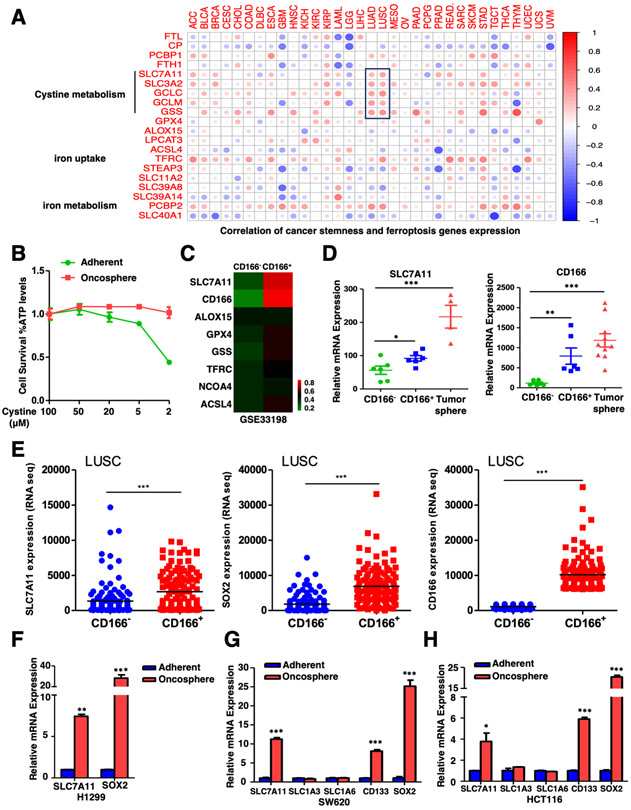
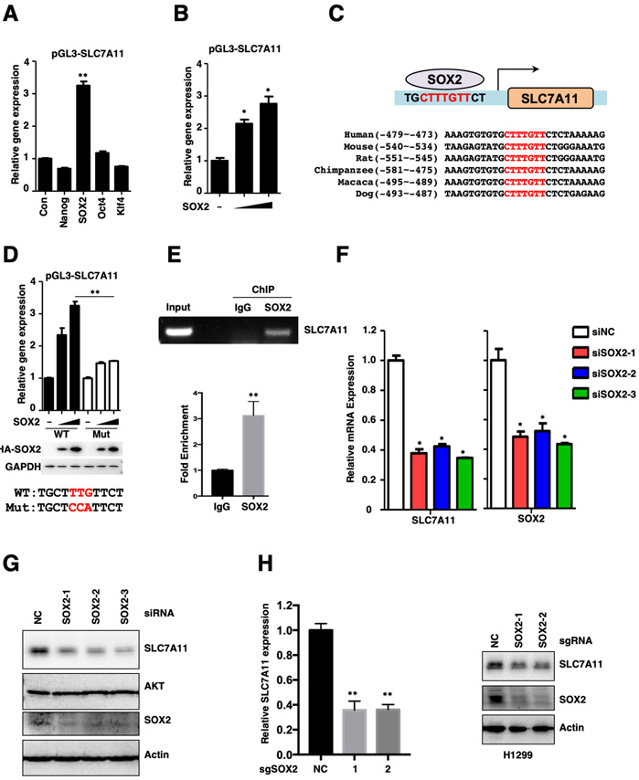
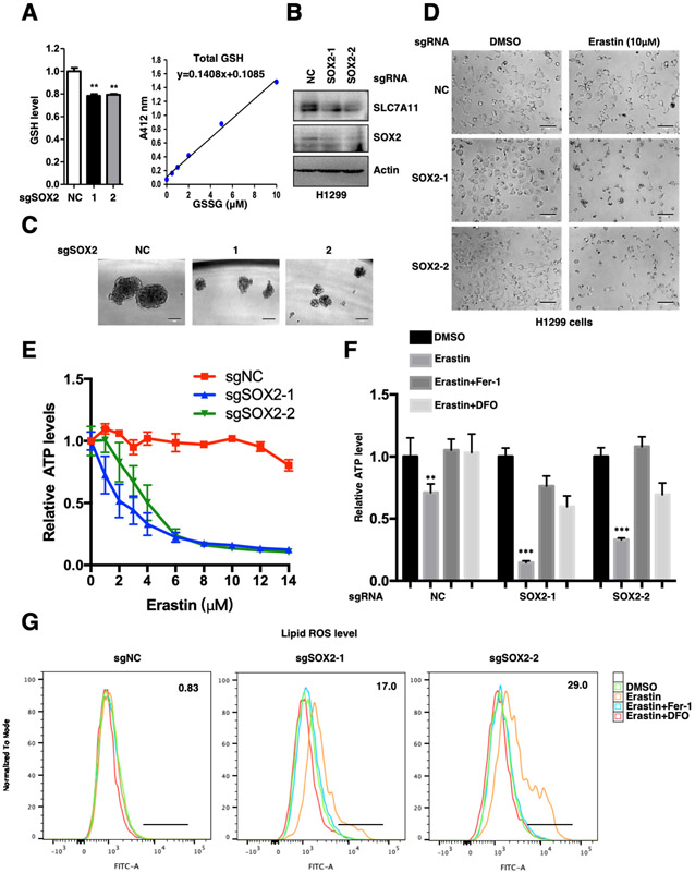
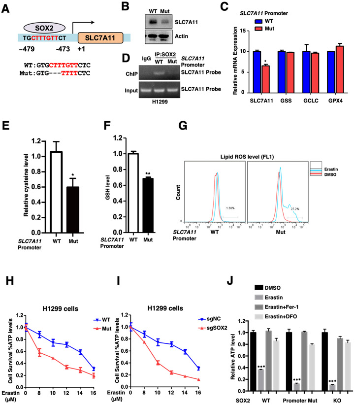
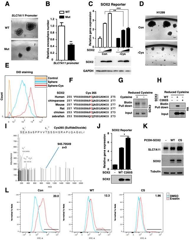
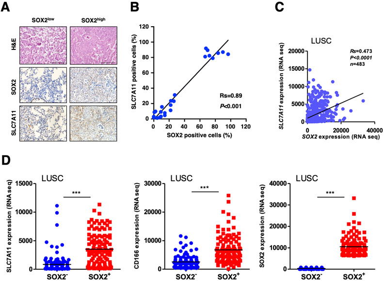
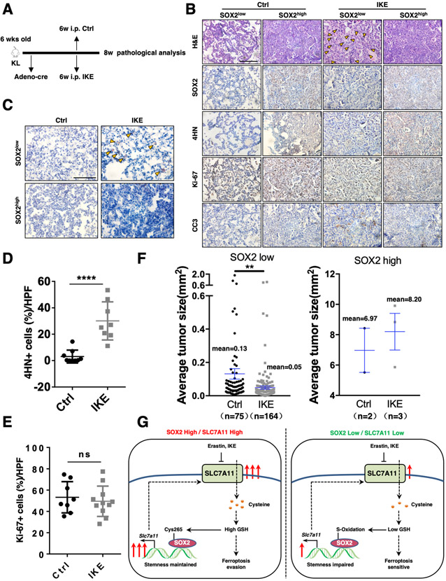
Ferroptosis is a lipid peroxidation-dependent cell death caused by metabolic dysfunction. Ferroptosis-associated enzymes are promising therapeutic targets for cancer treatment. However, such therapeutic strategies show limited efficacy due to drug resistance and other largely unknown underlying mechanisms. Here we report that cystine transporter SLC7A11 is upregulated in lung cancer stem-like cells (CSLC) and can be activated by stem cell transcriptional factor SOX2. Mutation of SOX2 binding site in SLC7A11 promoter reduced SLC7A11 expression and increased sensitivity to ferroptosis in cancer cells. Oxidation at Cys265 of SOX2 inhibited its activity and decreased the self-renewal capacity of CSLCs. Moreover, tumors with high SOX2 expression were more resistant to ferroptosis, and SLC7A11 expression was positively correlated with SOX2 in both mouse and human lung cancer tissue. Together, our study provides a mechanism by which cancer cells evade ferroptosis and suggests that oxidation of SOX2 can be a potential therapeutic target for cancer treatment. SIGNIFICANCE: This study uncovers a SOX2-SLC7A11 regulatory axis that confers resistance to ferroptosis in lung cancer stem-like cells.


Conflict of interest statement

CONFLICTS OF INTEREST STATEMENT:

The authors declare no potential conflicts of interest.

Figures

Figure.1
Figure.1. Cystine transporter SLC7A11 is upregulated in CSLCs
A. Correlation between cancer stemness and ferroptosis genes expression in 33 tumor types was shown. The Kruskal test in R was used to test the correlation, P value < 0.05 was considered statistically significant. ACC (Adrenocortical Carcinoma), BLCA (Bladder Urothelial Carcinoma), BRCA (Breast Invasive Carcinoma), CESC (Cervical Squamous Cell Carcinoma and Endocervical Adenocarcinoma), CHOL (Cholangiocarcinoma), COAD (Colon Adenocarcinoma), DLBC (Lymphoid Neoplasm Diffuse Large B-cell Lymphoma), ESCA (Esophageal Carcinoma), GBM (Glioblastoma Multiforme), HNSC (Head and Neck Squamous Cell Carcinoma), KICH (Kidney Chromophobe), KIRC (Kidney Renal Clear Cell Carcinoma), KIRP (Kidney Renal Papillary Cell Carcinoma), LAML (Acute Myeloid Leukemia), LGG (Brain Lower Grade Glioma), LIHC (Liver Hepatocellular Carcinoma), LUAD (Lung Adenocarcinoma), LUSC (Lung Squamous Cell Carcinoma), MESO (Mesothelioma), OV (Ovarian Serous Cystadenocarcinoma), PAAD (Pancreatic Adenocarcinoma), PCPG (Pheochromocytoma and Paraganglioma), PRAD (Prostate Adenocarcinoma), READ (Rectum Adenocarcinoma), SARC (Sarcoma), SKCM (Skin Cutaneous Melanoma), STAD (Stomach Adenocarcinoma), TGCT (Testicular Germ Cell Tumors), THCA (Thyroid Carcinoma), THYM (Thymoma), UCEC (Uterine Corpus Endometrial Carcinoma), UCS (Uterine Carcinosarcoma) and UVM (Uveal Melanoma). B. H1299 adherent and oncosphere cells were cultured in different concentrations of cysteine for 24 hours and cell viability of indicated cells was measured. The data was representative of three independent experiments. C. Gene expression data of CD166 control and CD166+ lung CSLCs was obtained from GEO (GSE33198) and heatmap of ferroptosis related gene expression was shown. D. Expression levels of SLC7A11 and CD166 in CD166 control cells, CD166+ lung CSLCs and lung tumor sphere were showed, *P<0.05, **P<0.01, ***P<0.001 (Student's t-test). E. Expression levels of SLC7A11, SOX2 and CD166 in CD166 versus CD166+ lung tumor cells from TCGA database were showed as dot plots, The data represented as scatter dot plot and the line showed mean. Significance was assessed by the Wilcoxon test compared between the indicated two groups, ***P<0.001 (Student's t-test). F. SLC7A11 was upregulated in H1299 tumor oncospheres when compared with adherent cells, **P<0.01, ***P<0.001 (Student's t-test). G. SLC7A11 but not other transporters was upregulated in SW620 tumor oncospheres when compared with adherent cells, ***P<0.001 (Student's t-test). H. SLC7A11 but not other transporters was upregulated in HCT116 tumor oncospheres when compared with adherent cells, *P<0.05, ***P<0.001 (Student's t-test).
Figure.2
Figure.2. SOX2 transcriptionally activates the expression of SLC7A11
A. pGL3-SLC7A11 promoter reporter gene was co-expressed with Nanog, SOX2, Oct4 and Klf4 in HEK293T cells for 24 hours, and the expression of Luciferase was measured and normalized to Renilla, **P<0.01 (Student's t-test). B. pGL3-SLC7A11 promoter reporter gene was co-expressed with different dose of SOX2 in HEK293T cells for 24 hours, and the expression of Luciferase was measured and normalized to Renilla, *P<0.05 (Student's t-test). C. Schematic diagram of SOX2 binding site on SLC7A11 promoter and sequence homology across different species. D. WT and mutated pGL3-SLC7A11 promoter reporter genes were co-expressed with different dose of SOX2 in HEK293T cells for 24 hours, and the expression of Luciferase was measured and normalized to Renilla, **P<0.01 (two-way ANOVA test), the protein level of SOX2 was analyzed by WB. E. ChIP results of SOX2 binding on SLC7A11 promoter in indicated H1299 cells. F. siRNA mediated SOX2 knock down resulted in decreased expression of SLC7A11 in H1299 cells, mRNA expression level was analyzed by qRT-PCR, *P<0.05 (Student's t-test). G. siRNA mediated SOX2 knock down resulted in decreased expression of SLC7A11 in H1299 cells, protein levels of SLC7A11 and SOX2 were analyzed by WB (related to Fig. 2F). H. Both the mRNA and protein expression levels of SLC7A11 were decreased upon CRISPR mediated SOX2 KO in H1299 cells, mRNA expression level was analyzed by qRT-PCR, **P<0.01 (Student's t-test) and the protein level was analyzed by WB.
Figure.3
Figure.3. SOX2 protects lung cancer cells from ferroptosis
A. Relative GSH levels in SOX2 WT and KO H1299 cell lines were showed, all samples were normalized to cell number. **P<0.01 (Student's t-test). GSH assay standard curve line was also showed. B. Protein levels of SLC7A11 and SOX2 were analyzed by WB. C. Representative sphere images from each condition of H1299 cells. Scale bar: 100 μm. SOX2 KO severely impaired the self-renewal of tumor spheres. D. SOX2 WT and KO H1299 cell lines were treated with 10 μM Erastin for 20 hours and representative images from each condition were shown. Scale bar: 100 μm. E. Cell viability of indicated H1299 cells was measured. The data was representative of three independent experiments. F. Indicated H1299 cells were treated with 10 μM Erastin for 20 hours, 1 μM Fer-1 and 50 μM DFO treatment could rescue Erastin-induced cell death. Cell viability of indicated cells was measured, **P<0.01, ***P<0.001 (two-way ANOVA test). G. Lipid ROS levels of indicated H1299 cells treated with 5 μM Erastin for 10 hours. 1 μM Fer-1 and 50 μM DFO treatment could rescue Erastin-induced lipid peroxidation.
Figure.4
Figure.4. SLC7A11 promoter mutation cells are susceptible to ferroptosis
A. Schematic diagram of SLC7A11 promoter WT and mutant H1299 cells. B. The protein levels of SLC7A11 in SLC7A11 promoter WT and mutant H1299 cells were analyzed by WB. C. Relative mRNA expression levels of SLC7A11, GSS, GCLC and GPX4 in SLC7A11 promoter WT and mutant H1299 cells were analyzed by qRT-PCR, *P<0.05 (Student's t-test). SLC7A11 but not other ferroptosis related genes was decreased in SLC7A11 promoter mutation cells. D. ChIP results of SOX2 binding on SLC7A11 promoter in WT and mutant H1299 cells. The mutation largely abolished SOX2’s binding on the SLC7A11 promoter. E. Relative cysteine levels in SLC7A11 promoter WT and mutant H1299 cells were showed, *P<0.05 (Student's t-test). F. Relative GSH levels in SLC7A11 promoter WT and mutant H1299 cells were showed, **P<0.01 (Student's t-test). G. Lipid ROS levels of SLC7A11 promoter WT and mutation H1299 cells treated with 5 μM Erastin for 10 hours. H. SLC7A11 promoter WT and mutant H1299 cells were treated with different dose of Erastin for 20 hours and cell viability of indicated cells was measured. The data was representative of three independent experiments. I. SOX2 WT and KO H1299 cells were treated with different dose of Erastin for 20 hours and cell viability of indicated cells was measured. The data was representative of three independent experiments. J. Indicated H1299 cells were treated with 10 μM Erastin for 20 hours, 1 μM Fer-1 and 50 μM DFO treatment could rescue Erastin-induced cell death. Cell viability of indicated cells was measured, ***P<0.001 (two-way ANOVA test).
Figure.5
Figure.5. Cysteine deprivation induces SOX2-Cys265 oxidative modification
A. Representative sphere images derived from SLC7A11 promoter WT and mutant H1299 cells. Scale bar: 100 μm. B. Relative tumor spheres number derived from SLC7A11 promoter WT and mutant cells, **P < 0.01 versus WT (Student’s t test). C. SOX2 activity reporter gene was co-expressed with different dose of SOX2 in HEK293T cells for 24 hours, then the cells were treated with cysteine deprivation for 6 hours or not, and the expression of Luciferase was measured and normalized to Renilla, **P<0.01, ***P<0.001 (two-way ANOVA test). The protein levels were analyzed by WB. Cysteine deprivation had little effect on expression level of SOX2. D. Representative sphere images derived from H1299 cells treated with cysteine deprivation for 14 days or not, Scale bar: 100 μm. E. DiD staining assay was performed to test the self-renewal of H1299 CSLCs. DiD, the cell labeling dye which is quite stable in cells, will decrease upon cell division. CSLCs treated with cysteine deprivation showed slower cell renewal rate compared to that cultured in normal medium. F. Sequence alignment of SOX2 Cys265 across different species. G. H1299 cells were treated with cysteine deprivation for 6 hours or not, the cell lysate was incubated with maleimide-biotin to label reduced cysteine groups. Pull-down of biotin-labeled proteins was followed by WB. H. HA-SOX2 and C265S mutant were transfected in HEK293T cells for 24 hours, the cell lysate was incubated with maleimide-biotin to label reduced cysteine groups. Pull-down of biotin-labeled proteins was followed by WB. I. Tandem mass spectrometry spectrum of Glu-C-digested SOX2 fragment containing the oxidized cysteine residues (blue line) was shown. J. SOX2 activity reporter gene was co-expressed with SOX2 WT or C265S mutant plasmids in HEK293T cells for 24 hours, the expression of Luciferase was measured and normalized to Renilla, *P<0.05 (Student's t-test). The protein levels were analyzed by WB. K. The protein levels of SLC7A11 and SOX2 in indicated H1299 cells were analyzed by WB. L. Lipid ROS levels of indicated H1299 cells treated with 5 μM Erastin for 10 hours.
Figure.6
Figure.6. SLC7A11 expression positively correlates with SOX2 in lung cancer
A. Representative images of SLC7A11 and SOX2 immunohistochemical staining in SOX2 high and low mouse lung tumor specimens. Scale bar: 100 μm. B. The correlation of SLC7A11 and SOX2 level was showed in dot plots in lung tumor lesions from KL mice. Gene expression correlation was assessed by the Spearman test between the indicated two groups. We counted SOX2 and SLC7A11 positive cells per high-power fields (HPF), respectively. C. The gene expression correlation of SLC7A11 and SOX2 was showed in dot plots in lung squamous cell carcinoma (LUSC). Gene expression correlation was assessed by the Spearman test between the indicated two groups. D. The mRNA expression levels of SLC7A11, SOX2 and CD166 were compared using scatter dot plots in SOX2low versus SOX2high lung squamous cell carcinoma. The data represented as scatter dot plots and the line showed mean. Significance was assessed by the Wilcoxon test compared between the indicated two groups, ***P<0.001.
Figure.7
Figure.7. SOX2 high lung tumors are more resistant to IKE treatment versus SOX2low tumors
A. The scheme of IKE treatment. The KL mice at 6 weeks post Ad-Cre infection were treated with IKE for 2 weeks, followed by tumor analysis. B. Representative H&E and IHC staining of SOX2, 4HN, Ki-67 and CC3 of Ad-Cre-infected KL mice from control and IKE groups, yellow arrowheads indicated lipid droplets. Scale bar: 100 μm. C. Oil Red O staining of IKE treated tumors showing lipid droplets (in red) of large size. Scale bar: 100 μm. D. Statistical analysis of the percentage of 4HN+ cells in the lung tumors control and IKE groups in Ad-Cre-infected KL mice. Data was shown as mean ± SEM. Significance was assessed by the Wilcoxon test compared between the indicated two groups, ****P<0.0001. IKE treatment induced higher levels of 4HN. E. Statistical analysis of the percentage of Ki-67+ cells in the lung tumors control and IKE groups in Ad-Cre-infected KL mice. Data was shown as mean ± SEM. Significance was assessed by the Wilcoxon test, ns, not significant. IKE treatment had little effect on Ki-67 levels. F. Quantification of individual tumor size of Ad-Cre-infected KL mice from control and IKE groups. Data was shown as mean ± SEM. Significance was assessed by the Wilcoxon test compared between the indicated two groups, **P<0.01. IKE treatment decreased the tumor size of SOX2 low but not SOX2 high tumors. G. Model of SOX2-SLC7A11 regulation axis.

References

    1. Dixon Scott J., Lemberg Kathryn M., Lamprecht Michael R., Skouta R, Zaitsev Eleina M., Gleason Caroline E., et al., Ferroptosis: An Iron-Dependent Form of Nonapoptotic Cell Death. Cell, 2012. 149(5): p. 1060–1072. - PMC - PubMed
    1. Zheng J and Conrad M, The Metabolic Underpinnings of Ferroptosis. Cell Metab, 2020. 32(6): p. 920–937. - PubMed
    1. Jiang X, Stockwell BR, and Conrad M, Ferroptosis: mechanisms, biology and role in disease. Nat Rev Mol Cell Biol, 2021. - PMC - PubMed
    1. Stockwell BR, Friedmann Angeli JP, Bayir H, Bush AI, Conrad M, Dixon SJ, et al., Ferroptosis: A Regulated Cell Death Nexus Linking Metabolism, Redox Biology, and Disease. Cell, 2017. 171(2): p. 273–285. - PMC - PubMed
    1. Green DR, The Coming Decade of Cell Death Research: Five Riddles. Cell, 2019. 177(5): p. 1094–1107. - PMC - PubMed

Publication types

MeSH terms