. 2021 Aug 17;118(33):e2102034118.
doi: 10.1073/pnas.2102034118.

HMGB1 released from nociceptors mediates inflammation

Affiliations

HMGB1 released from nociceptors mediates inflammation

Huan Yang et al. Proc Natl Acad Sci U S A. .

Abstract

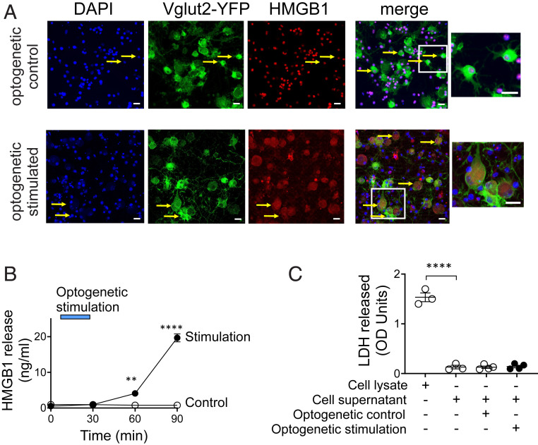
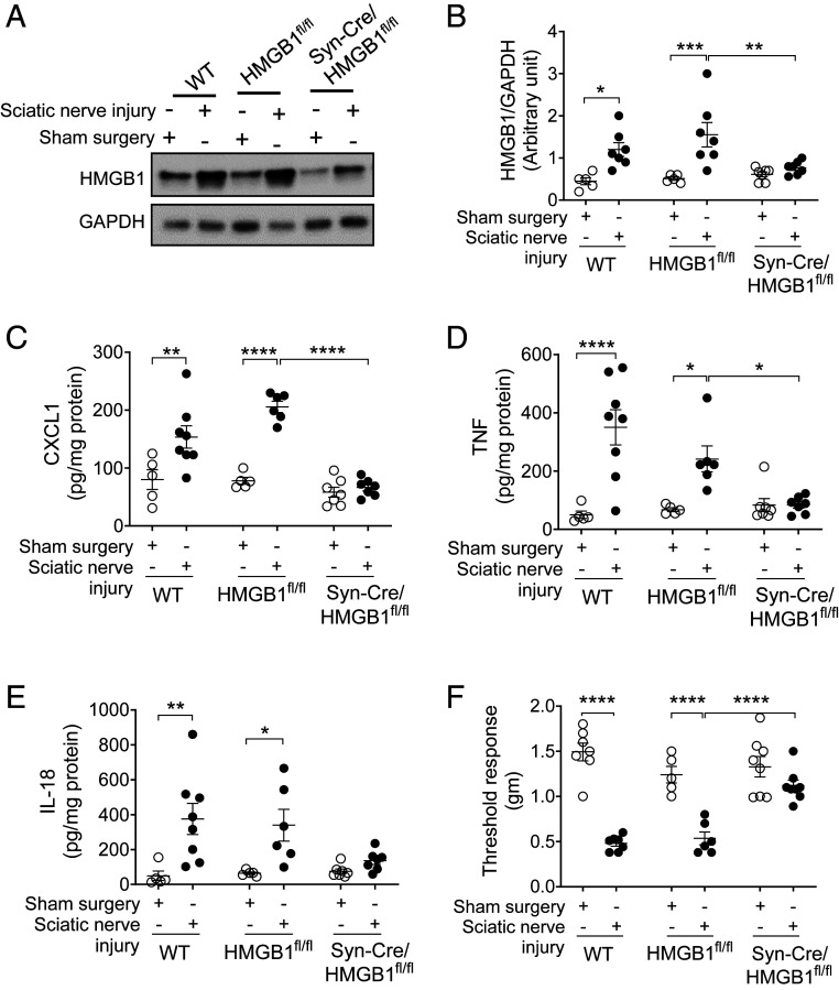
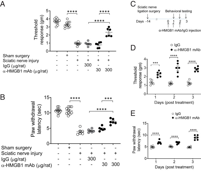
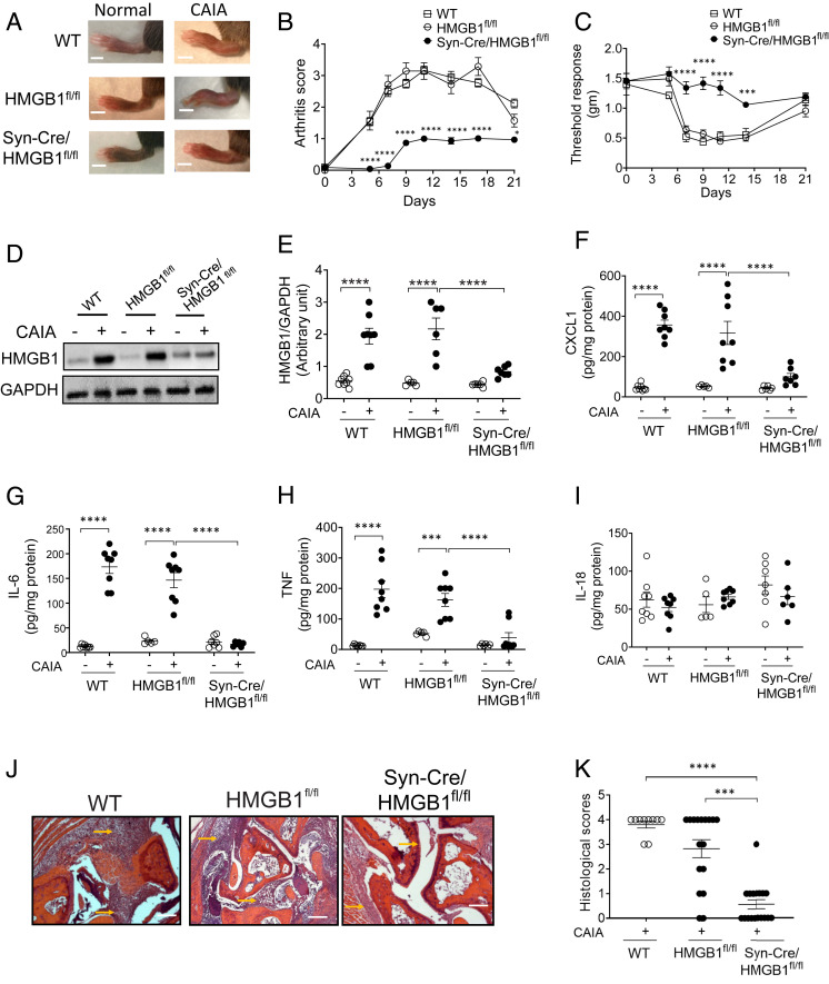
Inflammation, the body's primary defensive response system to injury and infection, is triggered by molecular signatures of microbes and tissue injury. These molecules also stimulate specialized sensory neurons, termed nociceptors. Activation of nociceptors mediates inflammation through antidromic release of neuropeptides into infected or injured tissue, producing neurogenic inflammation. Because HMGB1 is an important inflammatory mediator that is synthesized by neurons, we reasoned nociceptor release of HMGB1 might be a component of the neuroinflammatory response. In support of this possibility, we show here that transgenic nociceptors expressing channelrhodopsin-2 (ChR2) directly release HMGB1 in response to light stimulation. Additionally, HMGB1 expression in neurons was silenced by crossing synapsin-Cre (Syn-Cre) mice with floxed HMGB1 mice (HMGB1f/f). When these mice undergo sciatic nerve injury to activate neurogenic inflammation, they are protected from the development of cutaneous inflammation and allodynia as compared to wild-type controls. Syn-Cre/HMGB1fl/fl mice subjected to experimental collagen antibody-induced arthritis, a disease model in which nociceptor-dependent inflammation plays a significant pathological role, are protected from the development of allodynia and joint inflammation. Thus, nociceptor HMGB1 is required to mediate pain and inflammation during sciatic nerve injury and collagen antibody-induced arthritis.

Keywords: DAMP; HMGB1; arthritis; cytokine; sciatic nerve injury.


Conflict of interest statement

The authors declare no competing interest.

Figures

Fig. 1.
Fig. 1.
HMGB1 is actively released by stimulated sensory neurons. (A and B) Optogenetic stimulation of sensory neurons induces HMGB1 release. DRG neurons harvested from VGlut2-ChR2-YFP mice were stimulated with control yellow light (595 nm, n = 3) or blue light (470 nm, n = 4) at 20 Hz, 10% duty cycle for 30 min. (A) Confocal images showing translocation of HMGB1 from the nucleus to the cytoplasm. Fixed sensory neurons are stained with DAPI (blue), YFP (green, expressed in glutamatergic neurons), and anti-HMGB1 (red). The Inset shows the merged images of DAPI, YFP, and HMGB1 staining. Note the strong cytoplasmic translocation of HMGB1 in the sensory neurons stimulated with blue light, whereas neurons stimulated with control light exhibit predominant nuclear expression. (Scale bars, 20 µm.) Data are representative of three independent experiments. (B) Sensory neurons release HMGB1 upon activation. Blue bar indicates the duration of optogenetic stimulation. A time-dependent increase in secreted HMGB1 levels is observed following optogenetic stimulation of sensory neurons. Data are represented as mean ± SEM. Two-way ANOVA followed by Sidak’s multiple comparisons test between groups: control vs. blue light stimulation (at 60 min: **P < 0.01, at 90 min: ****P < 0.0001). (C) Optogenetic stimulation does not induce neuronal cell death. Cell viability was measured by LDH release. Cell lysate was included as a positive control for LDH measurements. n = 3 to 4 separate experiments, and each performed in duplicate. Data are represented as individual experimental data point with mean ± SEM. One-way ANOVA followed by Tukey’s multiple comparisons test between groups: ****P < 0.0001.
Fig. 2.
Fig. 2.
Neuronal HMGB1 ablation leads to reduced local inflammation and hyperalgesia after sciatic nerve injury. (A and B) Sciatic nerve injury induces increase in HMGB1 levels. (A) WT (n = 5 to 7), HMGB1fl/fl (n = 5 to 6), or Syn-Cre/HMGB1fl/fl mice (n = 7) were subjected to sciatic nerve ligation surgery or sham surgery. Representative images of Western blot for HMGB1 and GAPDH in the paw tissues 2 wk postsurgery. (B) Ratio of HMGB1/GAPDH expression. Data are represented as individual mouse data points with mean ± SEM. Two-way ANOVA followed by Sidak’s multiple comparisons test between groups: sham surgery vs. sciatic nerve injury (WT: *P < 0.05, HMGB1fl/fl: ***P < 0.001); sciatic nerve injury: HMGB1fl/fl vs. Syn-Cre/HMGB1fl/fl mice **P < 0.01. (C–E) Syn-Cre/HMGB1fl/fl mice mount reduced local chemokine and cytokines response. Levels of (C) CXCL1, (D) TNF, and (E) IL-18 in the paw tissues from WT (n = 5 to 8), HMGB1fl/fl (n = 5 to 6), or Syn-Cre/HMGB1fl/fl mice (n = 7). Data are represented as individual mouse data points with mean ± SEM. Two-way ANOVA followed by Sidak’s multiple comparisons test between groups: *P < 0.05, **P < 0.01, ****P < 0.0001. (F) Two weeks after sciatic nerve ligation surgery or sham surgery, mechanical hypersensitivity (von Frey) was assessed in WT (n = 7), HMGB1fl/fl (n = 5 to 6), or Syn-Cre/HMGB1fl/fl mice (n = 8). Data are represented as individual mouse data points with mean ± SEM. Two-way ANOVA followed by Sidak’s multiple comparisons test between groups: ****P < 0.0001.
Fig. 3.
Fig. 3.
Anti-HMGB1 mAb administration ameliorates sciatic nerve injury–induced hyperalgesia. (A and B) Female Wistar rats were subjected to sciatic nerve ligation surgery or sham surgery 2 wk prior to i.p. administration of anti-HMGB1 mAb (30 or 300 µg per rat) or control IgG (300 µg per rat). (A) Mechanical (von Frey) and (B) thermal (Hargreaves) hypersensitivity were assessed at 6 h after antibody was administered. Dose-dependent improvement in mechanical and thermal hypersensitivity is observed after anti-HMGB1 mAb administration. Data are represented as individual mouse data points with mean ± SEM. One-way ANOVA followed by Tukey’s multiple comparisons test between groups: ***P < 0.001, ****P < 0.0001. n = 10 per group (normal or sham) and n = 6 (sciatic nerve injury). (CE) Repetitive administration of anti-HMGB1 mAb ameliorates sciatic nerve injury–induced hyperalgesia. (C) Two weeks postsciatic nerve ligation or sham surgery, female Wistar rats received anti-HMGB1 mAb or IgG (300 µg/rat) i.p. once a day for 3 consecutive days. (D) Mechanical and (E) thermal hypersensitivity were assessed daily for 3 d after anti-HMGB1 mAb administration. Data are represented as individual mouse data points with mean ± SEM. Two-way ANOVA followed by Sidak’s multiple comparisons test between groups: ***P < 0.001, ****P < 0.0001, n = 6 per group.
Fig. 4.
Fig. 4.
Ablation of neuronal HMGB1 reduces joint inflammation and cartilage destruction and improves hyperalgesia in murine collagen antibody–induced arthritis. (A and B) Syn-Cre/HMGB1fl/fl mice are protected from CAIA. (A) Representative images of hind paws of WT, HMGB1fl/fl, and Syn-Cre/HMGB1fl/fl mice on day 10 after initiation of arthritis. (Scale bar, 0.2 cm.) (B) Arthritis scores are significantly reduced in Syn-Cre/HMGB1fl/fl mice as compared to WT and HMGB1fl/fl mice. Data are represented as mean ± SEM. Two-way ANOVA followed by Sidak’s multiple comparisons test between groups: Syn-Cre/HMGB1fl/fl vs. HMGB1fl/fl group: *P < 0.05, ****P < 0.0001. (C) Mechanical hypersensitivity (von Frey) is significantly improved in Syn-Cre/HMGB1fl/fl mice subjected to CAIA as compared to WT and HMGB1fl/fl mice. Data are represented as mean ± SEM. Two-way ANOVA followed by Sidak’s multiple comparisons test between groups: Syn-Cre/HMGB1fl/fl vs. HMGB1fl/fl group: ***P < 0.001, ****P < 0.0001. (D and E) HMGB1 levels are increased in the paw tissue in WT and HMGB1fl/fl mice but not in Syn-Cre/HMGB1fl/fl after CAIA. HMGB1 levels were assessed 10 d after initiation of arthritis in the paw tissues. (D) Representative images of Western blot for HMGB1 and GAPDH in the paw tissues post CAIA. (E) Ratio of HMGB1/GAPDH expression. Data are represented as individual mouse data points with mean ± SEM n = 8 (WT group), n = 5 to 6 (HMGB1fl/fl), and n = 6 to 7 (Syn-Cre/HMGB1fl/fl). Two-way ANOVA followed by Sidak’s multiple comparisons test between groups: ****P < 0.0001. (FI) Syn-Cre/HMGB1fl/fl mice mount reduced local chemokine and cytokines response. Levels of (F) CXCL1, (G) IL-6, (H) TNF, and (I) IL-18 in the paw tissue from WT (n = 8), HMGB1fl/fl (n = 5 to 8) or Syn-Cre/HMGB1fl/fl mice (n = 6 to 7). Data are represented as individual mouse data points with mean ± SEM. Two-way ANOVA followed by Sidak’s multiple comparisons test between groups: ***P < 0.001, ****P < 0.0001. (J and K) Histological assessment of joints on day 10 after initiation of arthritis. (J) Reduced infiltration of inflammatory cells, pannus formation, and cartilage damage (arrows) is observed in Syn-Cre/HMGB1fl/fl mice as compared to WT and HMGB1fl/fl subjected to CAIA. (Scale bars, 50 µm.) (K) The degree of inflammatory cells infiltration and cartilage disruption was graded as described in Materials and Methods. A significant reduction in histological score is observed in Syn-Cre/HMGB1fl/fl mice. n = 10 (WT), n = 18 (HMGB1fl/fl and Syn-Cre/HMGB1fl/fl). Data are represented as individual mouse data points with mean ± SEM. One-way ANOVA followed by Tukey’s multiple comparisons test between groups: ***P < 0.001, ****P < 0.0001.

Similar articles

Cited by

References

    1. Pearce J. M., Sir Charles Scott Sherrington (1857-1952) and the synapse. J. Neurol. Neurosurg. Psychiatry 75, 544 (2004). - PMC - PubMed
    1. Burke R. E., Sir Charles Sherrington’s the integrative action of the nervous system: a centenary appreciation. Brain 130, 887–894 (2007). - PubMed
    1. Pinho-Ribeiro F. A., W. A.Verri, Jr, Chiu I. M., Nociceptor sensory neuron-immune interactions in pain and inflammation. Trends Immunol. 38, 5–19 (2017). - PMC - PubMed
    1. Chavan S. S., Ma P., Chiu I. M., Neuro-immune interactions in inflammation and host defense: Implications for transplantation. Am. J. Transplant. 18, 556–563 (2018). - PMC - PubMed
    1. Silverman H. A., Chen A., Kravatz N. L., Chavan S. S., Chang E. H., Involvement of neural transient receptor potential channels in peripheral inflammation. Front. Immunol. 11, 2742 (2020). - PMC - PubMed

Publication types

MeSH terms

LinkOut - more resources