. 2021 Aug 17;118(33):e2108963118.
doi: 10.1073/pnas.2108963118.

A broadly active fucosyltransferase LmjFUT1 whose mitochondrial localization and activity are essential in parasitic Leishmania

Affiliations

A broadly active fucosyltransferase LmjFUT1 whose mitochondrial localization and activity are essential in parasitic Leishmania

Hongjie Guo et al. Proc Natl Acad Sci U S A. .

Abstract

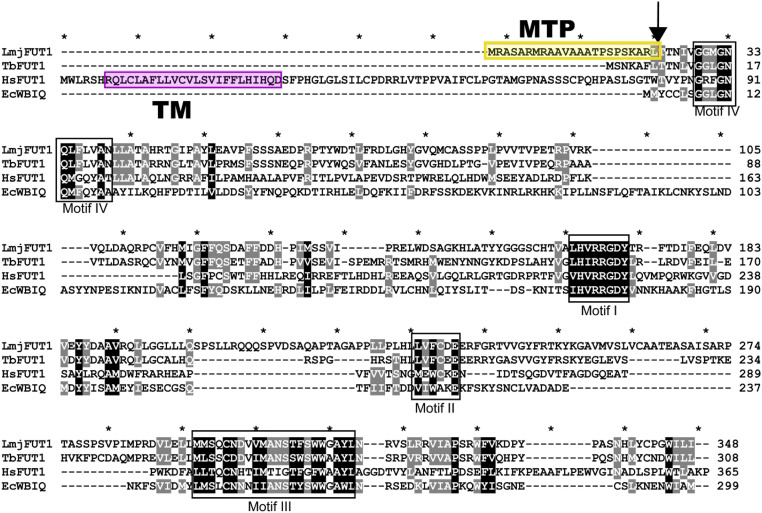
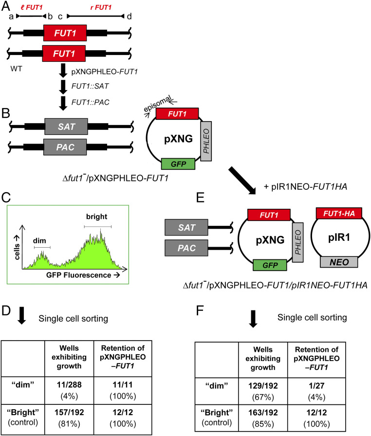
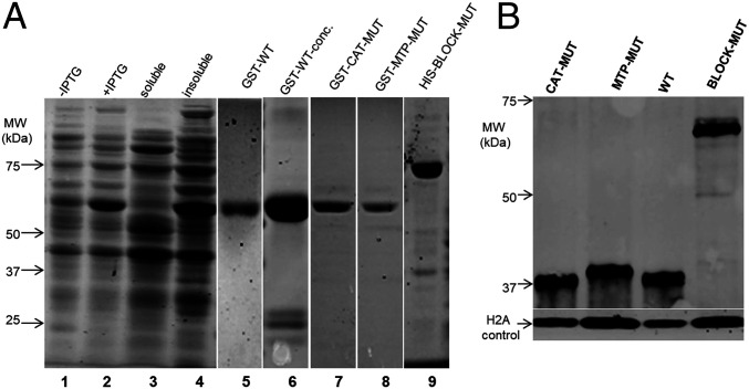
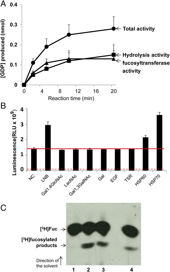
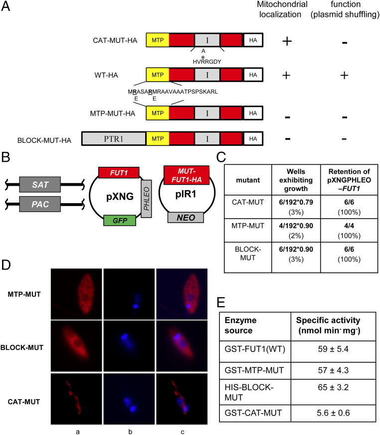
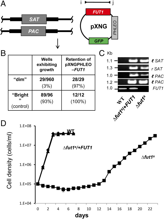
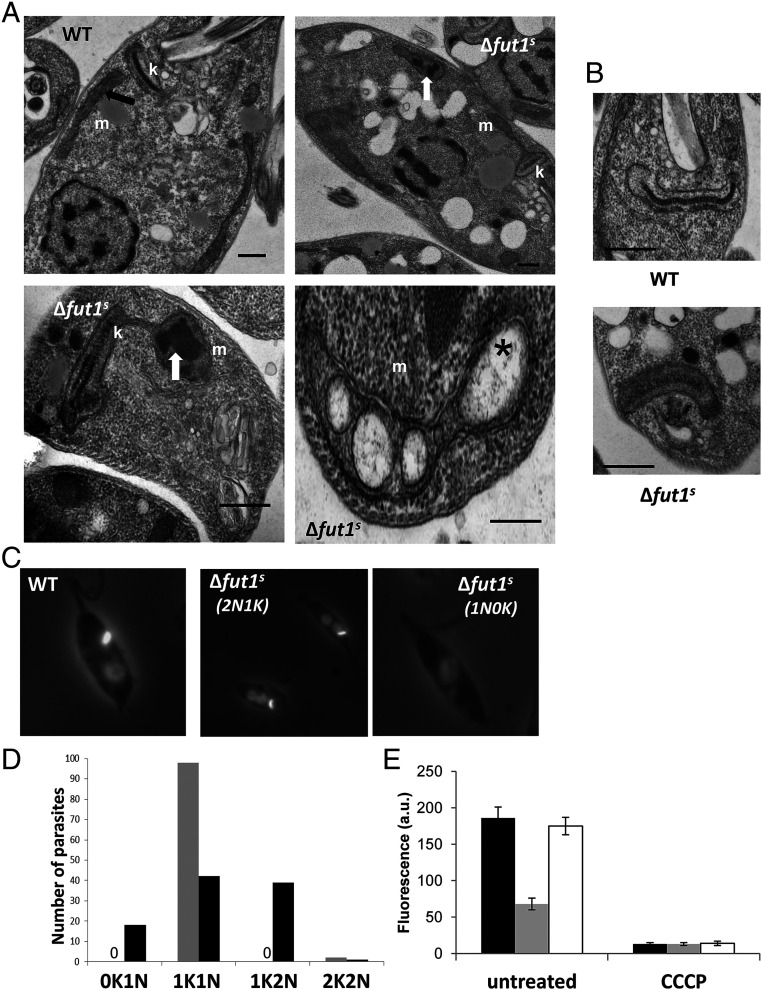
Glycoconjugates play major roles in the infectious cycle of the trypanosomatid parasite Leishmania While GDP-Fucose synthesis is essential, fucosylated glycoconjugates have not been reported in Leishmania major [H. Guo et al., J. Biol. Chem. 292, 10696-10708 (2017)]. Four predicted fucosyltransferases appear conventionally targeted to the secretory pathway; SCA1/2 play a role in side-chain modifications of lipophosphoglycan, while gene deletion studies here showed that FUT2 and SCAL were not essential. Unlike most eukaryotic glycosyltransferases, the predicted α 1-2 fucosyltransferase encoded by FUT1 localized to the mitochondrion. A quantitative "plasmid segregation" assay, expressing FUT1 from the multicopy episomal pXNG vector in a chromosomal null ∆fut1- background, established that FUT1 is essential. Similarly, "plasmid shuffling" confirmed that both enzymatic activity and mitochondrial localization were required for viability, comparing import-blocked or catalytically inactive enzymes, respectively. Enzymatic assays of tagged proteins expressed in vivo or of purified recombinant FUT1 showed it had a broad fucosyltransferase activity including glycan and peptide substrates. Unexpectedly, a single rare ∆fut1- segregant (∆fut1s ) was obtained in rich media, which showed severe growth defects accompanied by mitochondrial dysfunction and loss, all of which were restored upon FUT1 reexpression. Thus, FUT1 along with the similar Trypanosoma brucei enzyme TbFUT1 [G. Bandini et al., bioRxiv, https://www.biorxiv.org/content/10.1101/726117v2 (2021)] joins the eukaryotic O-GlcNAc transferase isoform as one of the few glycosyltransferases acting within the mitochondrion. Trypanosomatid mitochondrial FUT1s may offer a facile system for probing mitochondrial glycosylation in a simple setting, and their essentiality for normal growth and mitochondrial function renders it an attractive target for chemotherapy of these serious human pathogens.

Keywords: chemotherapy; fucose; glycobiology; glycosyltransferase; trypanosomatid protozoan parasites.


Conflict of interest statement

Competing interest statement: One of the reviewers (G.W.H.) and one of the authors (M.A.J.F.) are coauthors on chapter 12 in Essentials of Glycobiology (Cold Spring Harbor Laboratory Press, Cold Spring Harbor, NY, ed. 3, 2015–2017).

Figures

Fig. 1.
Fig. 1.
Comparison of mitochondrial Leishmania major FUT1 to fucosyltransferases from Trypanosoma brucei (TbFUT1), bacteria (EcWBIQ from E. coli O127), and mammals (human HsFUT1). Identical or highly conserved residues are highlighted in black or gray. Conserved fucosyltransferase motifs are marked by black boxes (40, 41). The predicted mitochondrial targeting peptide (MTP) is shaded in yellow, and cleavage site is marked by an arrow. The predicated transmembrane domain (TM) for HsFUT1 is shaded in pink.
Fig. 2.
Fig. 2.
Plasmid segregation tests show FUT1 is essential. (A) FUT1 locus and transfection. The figure shows the WT FUT1 locus, with the ORF as a central red box and the flanking sequences used for homologous replacement by SAT or PAC drug resistance cassettes (FUT1::SAT or FUT1::PAC, respectively) shown as heavier flanking lines. The location of “outside” primers used to establish planned replacements (a/b or c/d; to confirm replacements SAT or PAC specific primers were substituted for b or c; SI Appendix, Fig. S3) are shown. WT L. major was first transfected with pXNGPHLEO-FUT1, and successively with the FUT1::SAT and FUT::PAC constructs. (B) The chromosomal null Δfut1 mutant obtained in the presence of episomal FUT1. The genotype of this line is FUT::∆SAT/FUT1::∆PAC/ +pXNGPHLEO-FUT1, abbreviated as Δfut1/+pXNGPHLEO-FUT1. PCR confirmation tests of the predicted genotype are found in SI Appendix, Fig. S3. pXNG additionally expresses GFP (45). (C) Plasmid segregation tests of FUT1 essentiality. Δfut1/+pXNGPHLEO-FUT1 was grown for 24 h in the absence of phleomycin, and analyzed by GFP flow cytometry. The two gates used for subsequent quantitation and/or sorting of parasites are shown; dim or weakly fluorescent (2–10 FU) and bright or strongly fluorescent (100–1,000 FU). (D) Results of sorting experiment shown in C. Single cells from both GFP dim or bright populations sorted into 96-well plates containing M199 medium, and incubated for 2 wk, at which time growth was assessed. The numbers sorted and their growth and properties are shown, with bright cells mostly surviving and dim cells mostly not. All 11 survivors from the dim and 12 tested from the bright population sort were confirmed to retain of pXNGPHLEO-FUT1 by growth in media containing phleomycin. (E) Generation of line required for plasmid shuffling. Δfut1/+pXNGPHLEO-FUT1 was transfected with pIR1NEO-FUT1-HA expressing C-terminally tagged FUT1. PCR tests confirming the predicted genotype Δfut1/+pXNGPHLEO-FUT1/+pIR1NEO-FUT1-HA are shown in SI Appendix, Fig. S3. This line was then grown 24 h in the absence of phleomycin and subjected to cell sorting and growth tests as described in C. (F) Results of sorting experiment described in E. In this experiment, both dim and bright cells mostly survived (67% and 85%, respectively). Twenty-seven of the dim cells were tested, of which 26 had lost pXNGPHLEO-FUT1 by PCR tests, thereby representing the desired Δfut1/+pIR1NEO-FUT1-HA.
Fig. 3.
Fig. 3.
LmjFUT1 is localized to the parasite mitochondrion. (A) Indirect immunofluorescence of parasites lines of Δfut1/+pIR1NEO-FUT1-HA incubated with rabbit anti-HA antibody and visualized with goat anti-rabbit Alex 488-conjugated antibody (leftmost image, green), or MitoTracker Red CMXRos (central image, red), and merged (rightmost image, yellow) are shown. Colocalization by Pearson’s correlation coefficient was 0.97. (B) Cryo-immuno-EM of Δfut1/+pIR1NEO-FUT1-HA. FUT1-HA was visualized using rabbit anti-HA followed by gold-bead–conjugated mouse anti-rabbit IgG. F, flagellar pocket; K, kinetoplast DNA network; M, mitochondrion. Controls in which the primary anti-HA antibody was omitted yielded no bead counts. (C) Quantitation of cryo-immuno-EM anti-HA bead labeling to FUT-HA in cellular compartments. The data shown are the bead counts and SD, taken from three different experiments comprising three sets of 10 sections each, and normalized to the relative compartment area.
Fig. 4.
Fig. 4.
Expression of WT or mutant FUT1s in E. coli or within Leishmania. (A) Expression and purification of recombinant GST-FUT1 fusion proteins from E. coli visualized following SDS-PAGE. Lanes 1–6 are GST-FUT1 (WT). Lane 1: before-induction whole-cell lysates; lane 2: postinduction whole-cell lysates; lane 3: soluble fraction; lane 4: insoluble fraction; lane 5: elution; lane 6: concentrated eluted protein; lane 7, purified GST-CAT-MUT; lane 8, purified GST-MTP-MUT; and lane 9, purified HIS-BLOCK-MUT. Molecular weight markers are shown on the left. The images from lanes 1–4 and 5–9 are from separate experiments. (B) Western blot analysis of C-terminal HA-tagged FUT1s expressed in Leishmania. Lysates from parasites expressing the indicated HA-tagged FUT1s in a ∆fut1/+pXNGPHLEO-FUT1 background are shown (the presence of untagged WT FUT1 was required as none of the mutants were viable in its absence; Fig. 6). Western blots were performed with anti-HA to visualize the tagged FUT1 expression, and anti–L. major H2A as a loading control.
Fig. 5.
Fig. 5.
LmjFUT1 is a fucosyltransferase. (A) Time course of GDP production with GST-FUT1 in the presence or absence of GDP-Fucose and 1 mM LNB. Solid circles, incubation in the presence of GDP-Fucose + LNB (“total activity”); solid squares, incubation with GDP-Fucose alone (hydrolysis activity); solid triangles, difference between presence and absence of LNB (acceptor-dependent fucosyltransferase activity). (B) GST-FUT1 was incubated with GDP-Fucose in the presence of various acceptors, and the luminescence from GDP produced was measured after 5 min of incubation. Lane 1, NC, no acceptor (hydrolysis only); lane 2, LNB; lane 3, Gal1,4GlcNAc; lane 4, LacNAc; lane 5, Gal1,3GalNAc; lane 6; galactose; lane 7, EGF, EGF-like repeat; lane 8, thrombospondin like repeat; lane 9, mHSP60 peptide; and lane 10, mHSP70 peptide. The red horizontal line represents the hydrolysis activity (lane 1). The solid red line shows activity in the absence of acceptor or with inactive acceptors; the dashed red line marks the background seen in the absence of enzyme (about 7 × 10⁷ RLU under these conditions vs. 1.3 × 10⁹ RLU in the presence of enzyme). (C) Fucosyltransferase activity assayed in L. major in vivo. Whole-cell lysates were incubated with anti-HA agarose beads, which were washed, and then incubated with 1 μCi GDP-[3H]Fuc and 1 mM LNB as acceptor. Reaction products were separated by HPTLC and detected by fluorography as shown. Lane 1, WT; lane 2, Δfut1/+pIR1NEO-LmjFUT1-HA; lane 3, Δfut1/+pIR1NEO-TbFUT1; lane 4, WT/+pIR1NEO-Lmj-FUT1-HA. Elsewhere, we show that the level of free [3H]Fuc is considerably lower in the absence of enzyme than in its presence (46).
Fig. 6.
Fig. 6.
Both mitochondrial localization and fucosyltransferase activity are required for the essential function of FUT1. (A) Depiction of mutant LmjFUT1-HA designed to block mitochondrial import or catalysis. The predicted MTP is shown in yellow, the catalytic motif I is shown in gray, and the HA tag is shown in white, with the remainder of FUT1 in red. CAT-MUT-HA has an R297A substitution; MTP-MUT-HA replaces two Glu residues in the MTP with Arg; BLOCK-MUT-HA has the cytoplasmic protein PTR1 fused to the N terminus. The results of mitochondrial localization tests (D) and plasmid shuffling tests (C) are summarized on the Right of the illustrations. (B) Scheme of plasmid shuffling to test the function of mutant FUT1s. HA-tagged mutant FUT1s were expressed from pIR1NEO in the Δfut1/+pXNGPHLEO-FUT1 line. Growth in the absence of phleomycin and FACS sorting of dim and bright cells was performed as in Fig. 2. (C) Plasmid shuffling tests of FUT1 mutants. Δfut1pXNG-FUT1/pIR-MUT-HA lines was grown for 24 h in the absence of phleomycin, and analyzed by GFP flow cytometry. In all tests, survival of bright (control) cells was 79–90%. The results show that few dim cells yielded growth, and all of those arose from incomplete sorting (retention of pXNGPHLEO-FUT1). (D) Indirect immunofluorescence of HA-tagged FUT1 expressed from pIRNEO in the Δfut1/+pXNGPHLEO-FUT1 background. Column a, anti-HA (red); column b, DNA (Hoechst, blue); and column c, merge of columns a and b. (E) Acceptor-dependent specific activity of purified recombinant FUT1 proteins assayed by GDP formation in the presence of GDP-Fucose and LNB. The average and SD of three preparations are shown.
Fig. 7.
Fig. 7.
Recovery of a single FUT1-null mutant segregant (∆fut1s). (A) Starting cell line for plasmid segregation in rich culture medium. Δfut1/+pXNGPHLEO-FUT1 cells were grown and subjected to single-cell sorting in Schneider’s media. (B) Control bright cells showed a typically high frequency of wells supporting growth (93%), while few dim cells survived as in M199 medium (Fig. 2). Of the 29 survivors, 28 retained the pXNGPHLEO plasmid, while 1 designated ∆fut1s did not. (C) PCR tests of ∆fut1s, WT and a ∆fut1s /pXNGPHLEO-FUT1 “addback.” Primer sets l-SAT, r-SAT, l-PAC, and r-PAC confirm ∆fut1s lacks chromosomal FUT1 (SI Appendix, Fig. S3) and FUT1 ORF primers (l, 3955; j, 3956) confirm the absence of FUT1 sequences in ∆fut1s and its presence in the addback. (D) Growth of WT, ∆fut1s, and ∆fut1s/+pXNGPHLEO-FUT1 in M199 medium. Parasites were inoculated at a density of 105/mL and growth was followed by Coulter counting.
Fig. 8.
Fig. 8.
fut1s shows multiple mitochondrial abnormalities. (A) transmission EM of WT (subpanel a) and ∆fut1s (subpanels b–d). m, mitochondria; k, kinetoplast; black arrow, normal mitochondrial cristae; white arrows, aggregates inside mitochondria; star, bloated cristae. (Scale bars: 500 nm.) (B) Ultrastructural analysis of kDNA in WT and ∆fut1s. While WT cell presents typical compact kDNA structure, most mutant cells show a “looser” kDNA network that is wider with decreased length (see SI Appendix, Fig. S6 for quantitation). (Scale bar: 500 nm.) (C) Loss of kDNA network in ∆fut1s. WT or ∆fut1s were stained with DAPI to visualize the kDNA network (small bright spot) and nucleus (dimmer large circle). A typical one kinetoplast/one nucleus (1K/1N) pattern is shown for WT in the leftmost panel; the central panel shows a ∆fut1s cell with a 1K/2N pattern, and the rightmost panel shows a ∆fut1s cell lacking kDNA (0K/1N). (D) Quantitation of kDNA/nucleus patterns seen in WT (gray bars) vs. ∆fut1s (dark bars). A “0” indicates no cells of that pattern. (E) Mitochondrial potential assessed by staining with TMRE. Parasites were incubated with 100 nM TMRE and analyzed by flow cytometry, with the signal on the FL-2 channel expressed in arbitrary units (a.u.). The left portion shows cells incubated for 15 min; the right portion shows cells further incubated with 300 µM cyanide m-chlorophenylhydrazone (CCCP) for 60 min. Lines tested are as follows: WT, black bar; ∆fut1s, gray bar; and ∆fut1s/+pXNGPHLEO-FUT1, white bar.

References

    1. Alvar J.et al. .; WHO Leishmaniasis Control Team , Leishmaniasis worldwide and global estimates of its incidence. PLoS One 7, e35671 (2012). - PMC - PubMed
    1. Pigott D. M., et al. ., Global distribution maps of the leishmaniases. eLife 3, e02851 (2014). - PMC - PubMed
    1. Bañuls A. L., et al. ., Clinical pleiomorphism in human leishmaniases, with special mention of asymptomatic infection. Clin. Microbiol. Infect. 17, 1451–1461 (2011). - PubMed
    1. Naderer T., McConville M. J., Intracellular growth and pathogenesis of Leishmania parasites. Essays Biochem. 51, 81–95 (2011). - PubMed
    1. McConville M. J., Mullin K. A., Ilgoutz S. C., Teasdale R. D., Secretory pathway of trypanosomatid parasites. Microbiol. Mol. Biol. Rev. 66, 122–154 (2002). - PMC - PubMed

Publication types

MeSH terms