. 2021 Oct;16(10):1130-1140.
doi: 10.1038/s41565-021-00950-z. Epub 2021 Aug 12.

Immunogenic camptothesome nanovesicles comprising sphingomyelin-derived camptothecin bilayers for safe and synergistic cancer immunochemotherapy

Affiliations

Immunogenic camptothesome nanovesicles comprising sphingomyelin-derived camptothecin bilayers for safe and synergistic cancer immunochemotherapy

Zhiren Wang et al. Nat Nanotechnol. 2021 Oct.

Abstract

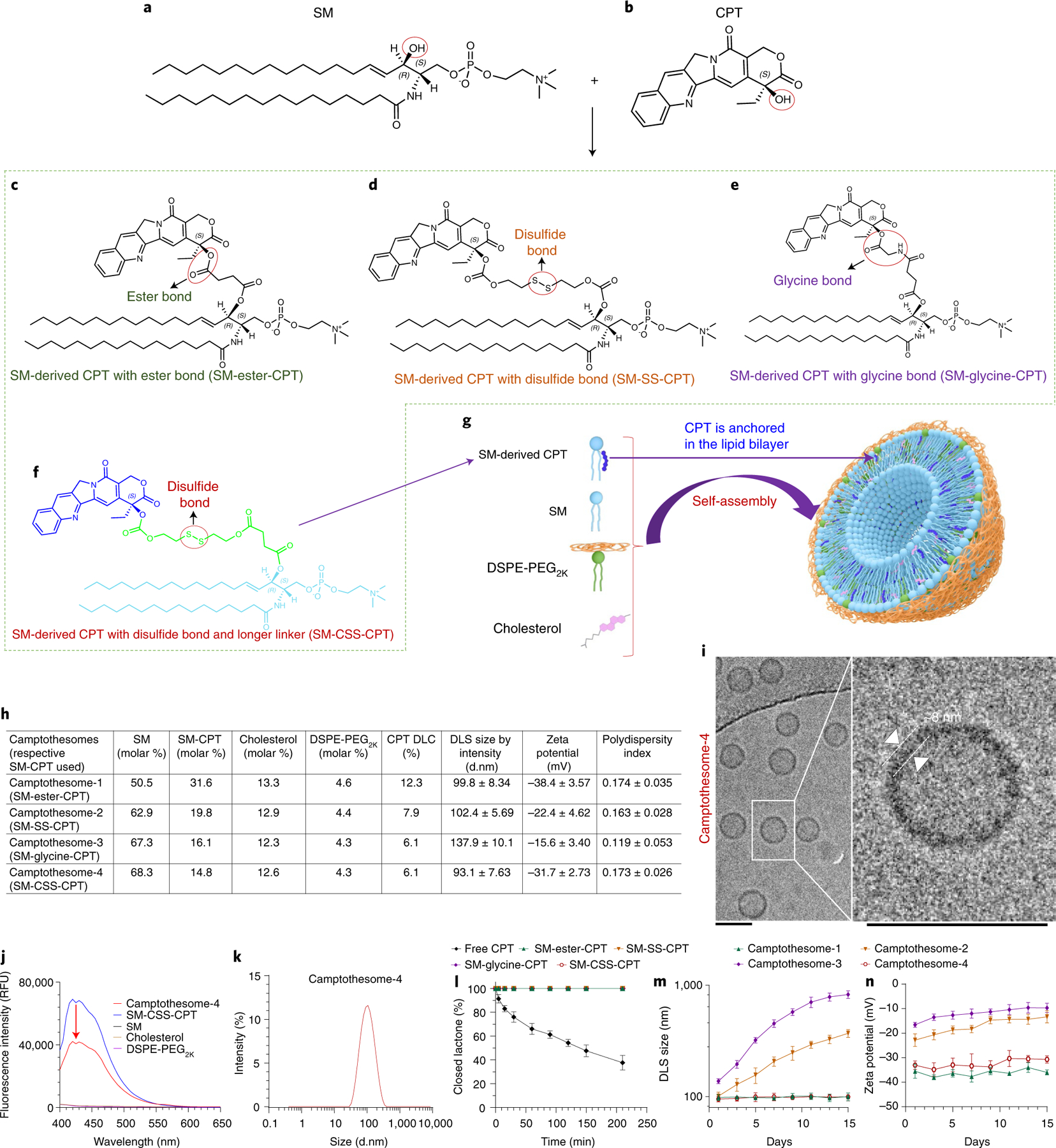
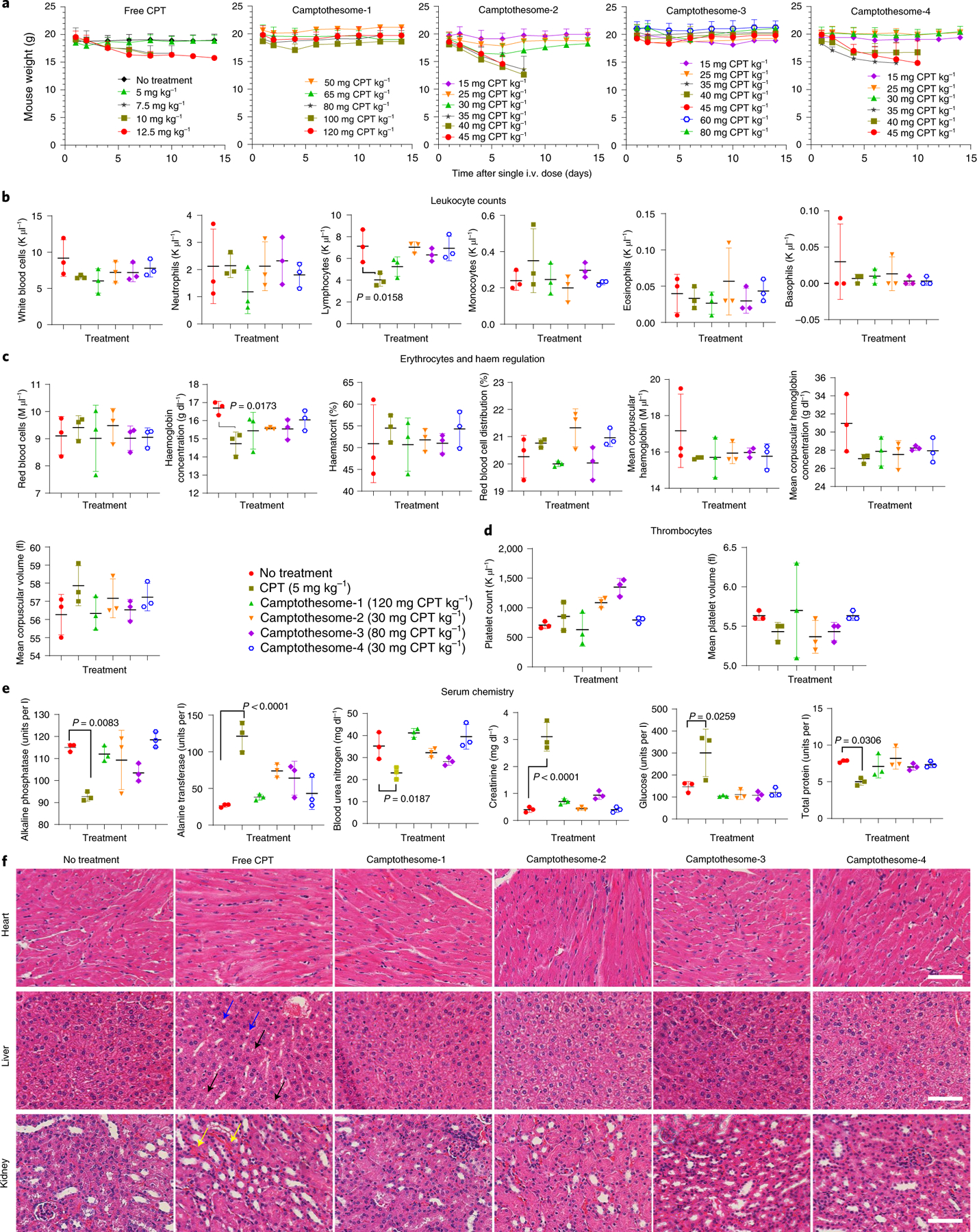
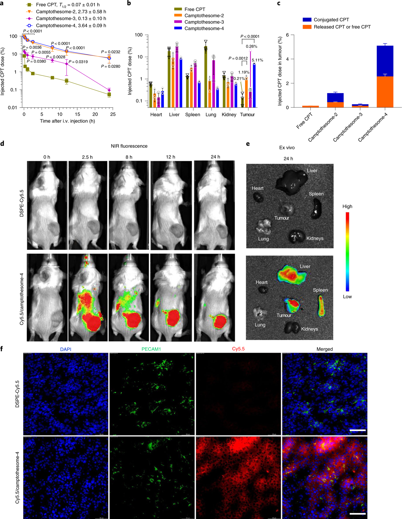
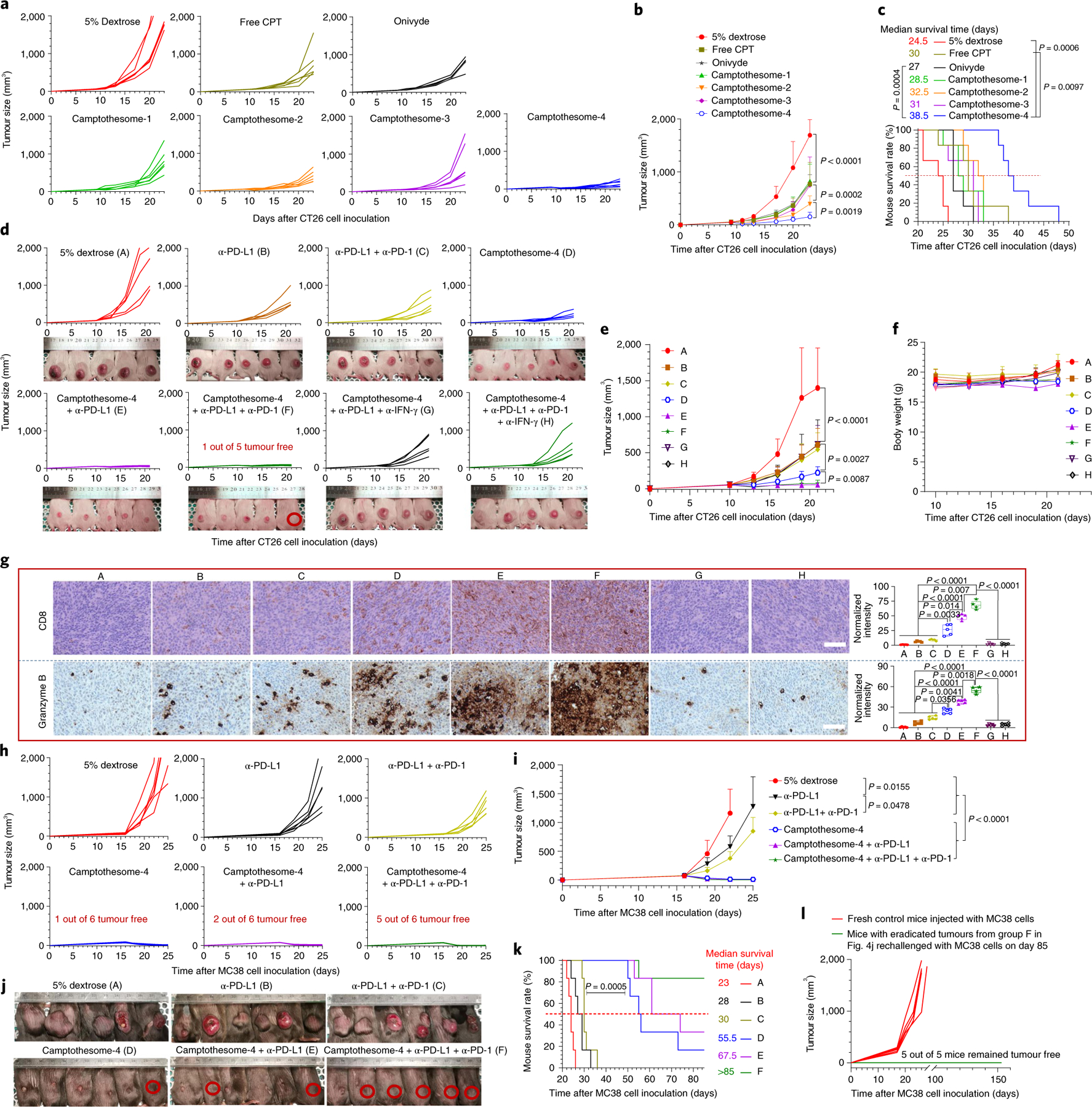
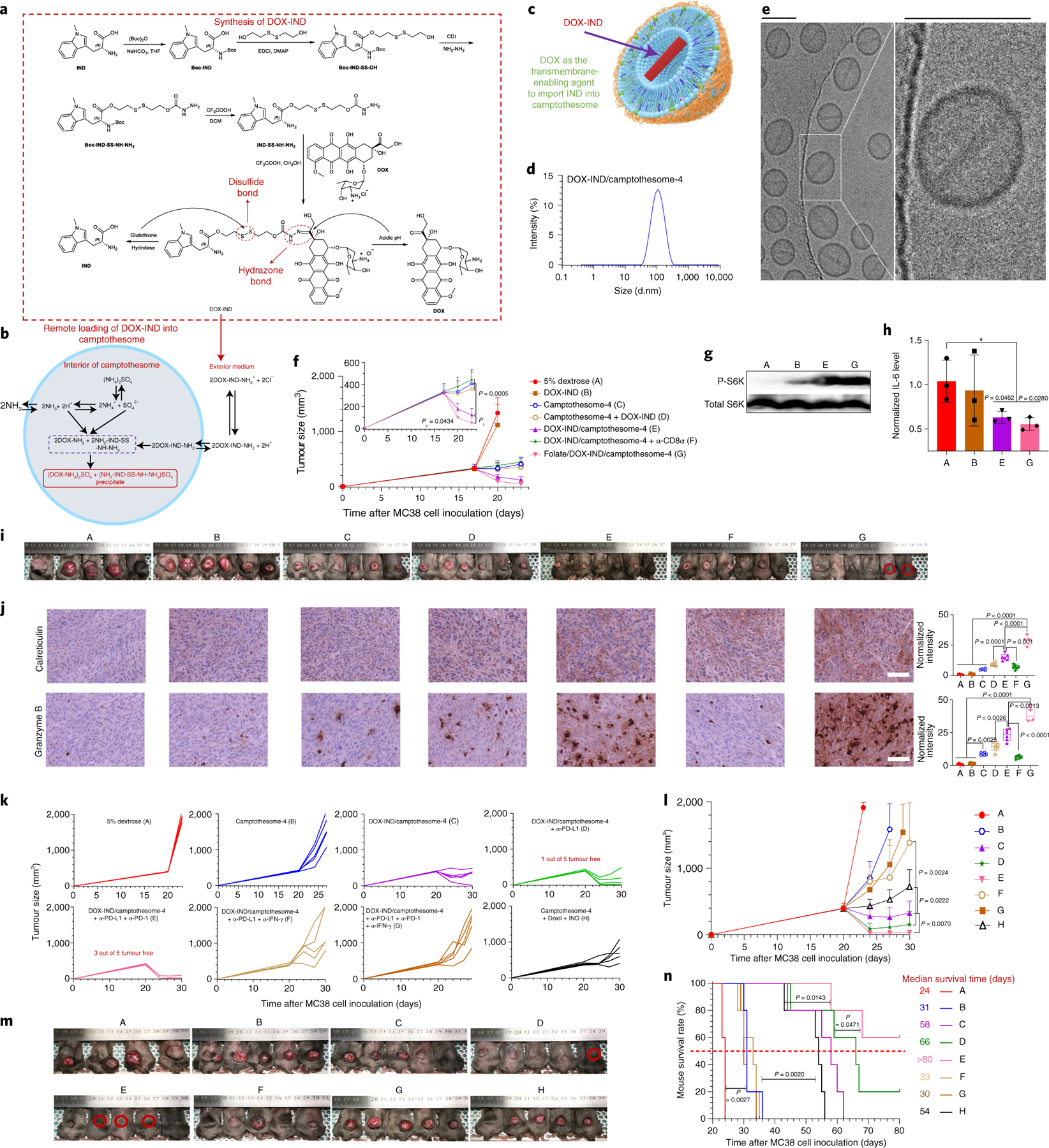
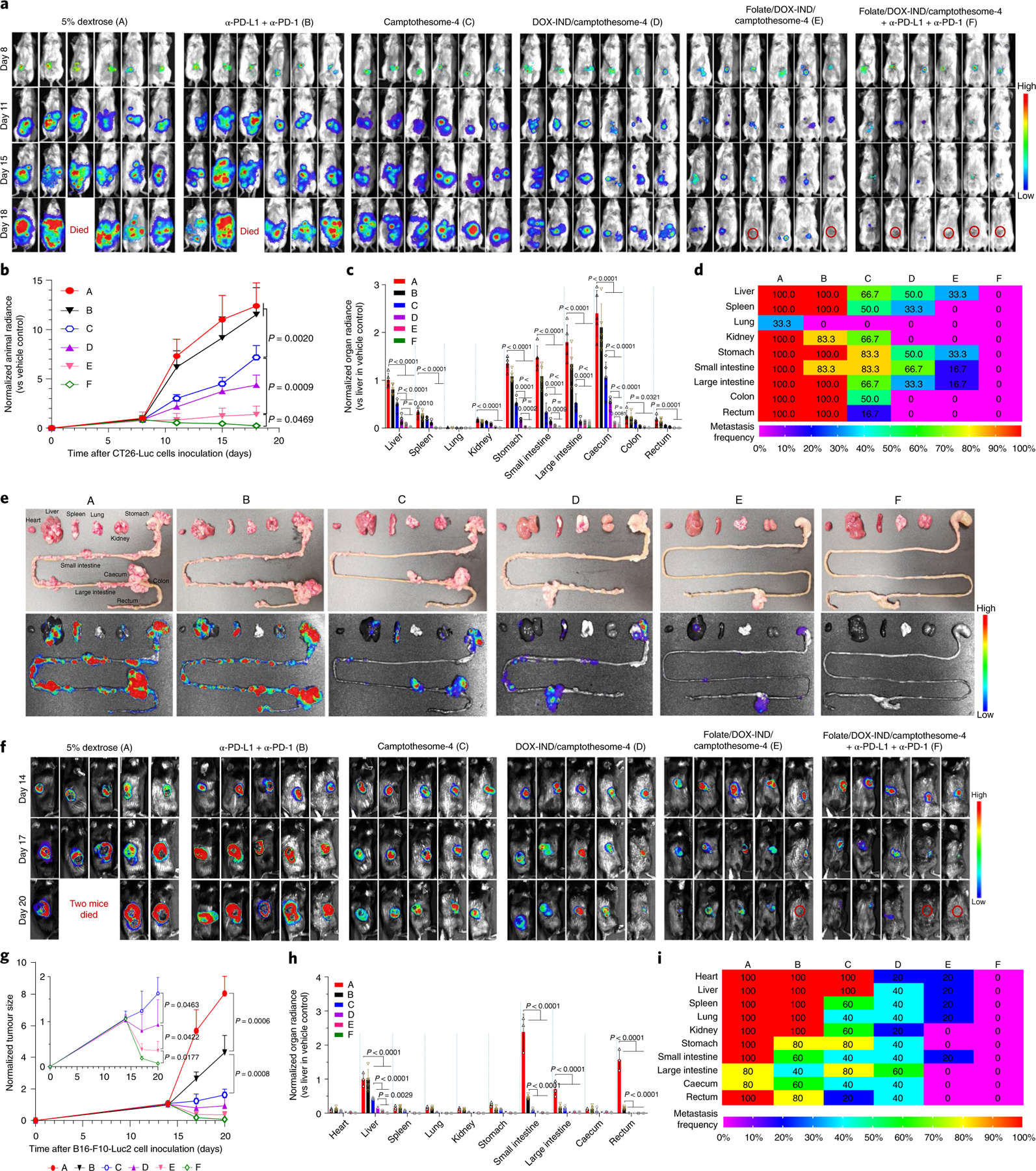
Despite the enormous therapeutic potential of immune checkpoint blockade (ICB), it benefits only a small subset of patients. Some chemotherapeutics can switch 'immune-cold' tumours to 'immune-hot' to synergize with ICB. However, safe and universal therapeutic platforms implementing such immune effects remain scarce. We demonstrate that sphingomyelin-derived camptothecin nanovesicles (camptothesomes) elicit potent granzyme-B- and perforin-mediated cytotoxic T lymphocyte (CTL) responses, potentiating PD-L1/PD-1 co-blockade to eradicate subcutaneous MC38 adenocarcinoma with developed memory immunity. In addition, camptothesomes improve the pharmacokinetics and lactone stability of camptothecin, avoid systemic toxicities, penetrate deeply into the tumour and outperform the antitumour efficacy of Onivyde. Camptothesome co-load the indoleamine 2,3-dioxygenase inhibitor indoximod into its interior using the lipid-bilayer-crossing capability of the immunogenic cell death inducer doxorubicin, eliminating clinically relevant advanced orthotopic CT26-Luc tumours and late-stage B16-F10-Luc2 melanoma, and achieving complete metastasis remission when combined with ICB and folate targeting. The sphingomyelin-derived nanotherapeutic platform and doxorubicin-enabled transmembrane transporting technology are generalizable to various therapeutics, paving the way for transformation of the cancer immunochemotherapy paradigm.


Conflict of interest statement

Competing interests

J.L. has applied for patents related to this study. The other authors have no competing interests.

Figures

Fig. 1 |
Fig. 1 |. Development of SM-derived CPT liposomal nanovesicles (camptothesomes).
a,b, Chemical structures of SM (a) and CPT (b). cf, Conjugation of SM and CPT resulted in SM-derived CPTs with either an ester bond (SM-ester-CPT) (c), a disulfide linkage (SM-SS-CPT) (d), a glycine bond (SM-glycine-CPT) (e), or a disulfide linkage and a longer linker (SM-CSS-CPT) (f). g, Schematic depicting the self-assembly of SM-CPT into a camptothesome. h, A table delineating the physicochemical characterizations of the four camptothesomes. DLS, dynamic light scattering; d.nm, diameter values in nanometres. i, cryo-EM for camptothesome-4. Scale bars, 100 nm. j, The fluorescence intensities (relative fluorescence units, RFU) for camptothesome-4, SM-CSS-CPT, SM, cholesterol and DSPE-PEG2K in methanol at equivalent concentrations. The significant fluorescence quenching for SM-CSS-CPT after self-assembly into camptothesome-4 demonstrates that there are strong π–π stacking interactions among SM-CSS-CPT molecules since CPT contains several aromatic rings. k, Representative DLS size distribution by intensity for camptothesome-4. For i–k, three independent experiments were performed to confirm the results. l, Percent closed lactone in PBS (pH 7.4) as a function of time for free CPT and the four different SM-CPT conjugates. SM-conjugated CPTs strongly prevented the lactone form from being converted into the inactive carboxylate form, enhancing CPT stability. m,n, Monitoring of the DLS size by intensity (m) and zeta potential (n) for each of the four different camptothesomes over time in 5% dextrose at 4 °C. Data in h (right portion) and ln are expressed as mean ± s.d. (n = 3 independent experiments).
Fig. 2 |
Fig. 2 |. Camptothesomes increased the MTD of CPT without inducing systemic toxicities in healthy mice.
a, Weight changes in healthy BALB/c mice in a MTD study of free CPT and the four camptothesomes administered intravenously once at various doses (as indicated) via a tail vein. Mouse body weight and survival were monitored for 2 weeks. The MTD was defined as the dose that did not cause mouse death or a weight loss of more than 15% during the whole period,,. Weight monitoring was terminated for the mice in the same dose group when there was any mouse death. bf, On day 14 post i.v. injection, blood was withdrawn for leukocytes (b), erythrocytes (c), thrombocytes (d) and serum chemistry (e) analysis, and the heart, liver (blue arrow, hepatic steatosis; black arrow, diffuse microvesicular degeneration of hepatocytes), and kidney (yellow arrow, haemorrhage in interstitial tissue) were isolated from the mice in the MTD groups (free CPT, 5 mg kg−1; camptothesome-1, 120 mg CPT kg−1; camptothesome-2, 30 mg CPT kg−1; camptothesome-3, 80 mg CPT kg−1; and camptothesome-4, 30 mg CPT kg−1) and the no-treatment group for haematoxylin and eosin (H&E) staining (f). Scale bar, 100 μm. Data in ae are expressed as mean ± s.d. (n = 3). For H&E staining, representative samples were taken from three independent mice in each group. Statistical significance was determined by one-way ANOVA followed by Tukey’s multiple comparisons test.
Fig. 3 |
Fig. 3 |. Improved circulation half-life and tumour delivery with efficient intratumoral drug release and deep tumour penetration.
a, Blood kinetics of CPT in subcutaneous CT26 tumour-bearing mice (n = 3 mice; tumours, ~300 mm3) following single i.v. injection of camptothesomes (20 mg CPT kg−1) and free CPT (5 mg kg−1, MTD). b,c, Tissue distribution (b) and CPT intratumoural release (c) at 24 h in mice in (a). The per cent injected doses in camptothesomes represent the released CPT and SM-conjugated CPT. Drug contents in plasma and major tissues were measured by HPLC (Supplementary Fig. 13). d,e, Real-time Lago optical imaging (d) and ex vivo imaging of different organs (e) from a representative mouse after single i.v. administration of free DSPE-Cy5.5 and DSPE-Cy5.5-labelled camptothesome-4 into subcutaneous CT26 tumour-bearing BALB/c mice (n = 3 mice; tumours, ~300 mm3). f, Investigation of the ability of camptothesome-4 to extravasate and penetrate the tumour after i.v. administration into mice with subcutaneous CT26 tumours (n = 3 mice, tumours, ~300 mm3). 24 h after i.v. injection of Cy5.5/camptothesome-4 (red), confocal laser scanning microscopy of sections of CT26 tumours was performed. Blood vessels were marked with PECAM1 antibody followed by Alexa Fluor 488 secondary antibody staining (green). Cell nuclei were stained by DAPI (blue). Scale bars, 50 μm. The results in ac are expressed as mean ± s.d. Statistical significance was determined by one-way ANOVA followed by Tukey’s multiple comparisons test and compared to free CPT.
Fig. 4 |
Fig. 4 |. Camptothesome synergizes with PD-L1/PD-1 blockade to eradicate CRC tumours.
a, Individual tumour growth curves (TGCs) in subcutaneous CT26 tumour-bearing mice (n = 6 mice; tumours, ~50 mm3) receiving a single i.v. injection of free CPT (5 mg kg−1), camptothesome or Onivyde at 20 mg CPT or irinotecan per kg on day 9; 5% dextrose was the vehicle control. b, Average TGCs. c, Kaplan–Meier survival curves. d, Individual TGCs for CT26 tumour-bearing mice (n = 5) receiving a single i.v. dose of camptothesome-4 (20 mg CPT kg−1) alone or combined with i.p. α-PD-L1 or α-PD-L1/α-PD-1 (100 μg per mouse per 3 day on three occasions) with or without α-IFN-γ on or from the tenth day. α-IFN-γ was intraperitoneally injected (200 μg per mouse per 3 days). Mouse images were taken on day 21. Red circle, tumour-free mouse. e, Average TGCs. f, Mouse body weight. g, Immune phenotypic analysis of the tumours in d using immunohistochemistry (IHC). Labels A to H in g correspond to those in d. In the box-and-whisker plots (right panel), boxes delineate lower and upper quartiles, middle lines show median values, dots show individual points, and whiskers show minimal and maximal values for each dataset. Scale bar, 100 μm. h, Individual TGCs for MC38 tumour-bearing mice (n = 6 mice; tumours, ~50 mm3) receiving a single i.v. dose of camptothesome-4 (20 mg CPT kg−1). α-PD-L1 and α-PD-1 were used similarly. i, Average TGCs. jl, Tumour-bearing mouse images taken on day 26 (j, one mouse death from the vehicle control group on day 23); red circle, tumour-free mouse. Kaplan–Meier survival curves (k); five tumour-free mice from group F in j were rechallenged with MC38 cells on day 85 (l). Labels A to F in k correspond to those in j. Data in b, e, f, i are expressed as mean ± s.d. Statistical significance was determined by one-way ANOVA followed by Tukey’s multiple comparisons test; survival curves were compared using the log-rank Mantel–Cox test.
Fig. 5 |
Fig. 5 |. Co-encapsulating DOX-IND in camptothesome-4 using DOX as a transmembrane-enabling carrier.
a, Synthesis of DOX-IND. b, Schematic of the remote incorporation of IND into camptothesome utilizing DOX as a lipid membrane-crossing carrier with (NH4)2SO4 as a pH gradient. Once DOX-IND is inside camptothesome-4, the acidic pH produced by (NH4)2SO4 breaks the hydrazone bond, releasing free DOX and IND intermediate. The –NH2 groups from both DOX and IND-SS-NH-NH2 enable the formation of (DOX-NH3)2SO4 and (NH3-IND-SS-NH-NH3)SO4 aggregated salts with dissociated SO42−, avoiding drug leakage or escape. c, Illustration of the co-encapsulation of DOX-IND into camptothesome. d,e, Representative DLS size distribution (d) and cryo-EM (e) of DOX-IND/camptothesome-4 from three independent experiments. Scale bar, 100 nm. fj, Antitumour efficacy in subcutaneous MC38 tumour-bearing mice (n = 6 mice; tumours, ~300 mm3) receiving a single i.v. injection on day 17 at the equivalent of 20 mg CPT kg−1 and 6.7 mg DOX-IND kg−1. α-CD8α (monoclonal antibody neutralizes mouse CD8α, 200 μg per mouse per 3 days) was given from day 17 via i.p. injection. Shown are average TGCs (f), representative western blotting for P-S6K (g) and qRT-PCR for IL-6 (h) from three independent tumours. Also shown are mouse images on day 23 (i, one mouse death from the vehicle control group on day 22; red circle, tumour-free mouse), and representative IHC staining and semi-quantitative analysis for calreticulin and granzyme B from independent tumours in i (j). Labels A to G in g and i correspond to those in f. In the box-and-whisker plots (j, right panel), boxes delineate lower and upper quartiles, middle lines show median values, dots show individual data points, and whiskers show minimal and maximal values for each dataset). Scale bar, 100 μm. kn, Subcutaneous MC38 tumour-bearing mice (n = 5 mice; tumours, ~400 mm3) received a single i.v. injection on day 20 at the equivalent of 15 mg CPT kg−1 and 5 mg DOX-IND kg−1. α-PD-L1, α-PD-1 and α-IFN-γ were administered via i.p. injection as described above. Individual TGCs (k), average TGCs (l), mouse images on day 24 (m, one mouse death from vehicle control group on day 21; red circle, tumour-free mouse) and Kaplan–Meier survival curves (n). Labels A to H in m and n correspond to those in l. Data in f, h, l are presented as mean ± s.d. Statistical significance was determined by one-way ANOVA followed by Tukey’s multiple comparisons test; survival curves were compared using the log-rank Mantel–Cox test.
Fig. 6 |
Fig. 6 |. Eradication of advanced and metastatic orthotopic CRC and subcutaneous melanoma tumours.
ae, Therapeutic efficacy in orthotopic CRC tumour mouse model. Mice were inoculated with 2 × 10⁶ CT26-Luc cells (RPMI-1640/Matrigel, 3/1, v/v) into the caecum subserosa. On day 8, mice (n = 6 mice; tumours, ~300 mg) were intravenously administered one dose of camptothesome-4, DOX-IND/camptothesome-4 or folate/DOX-IND/camptothesome-4 at the equivalent of 15 mg CPT kg−1 and 5 mg DOX-IND kg−1. α-PD-L1 and α-PD-1 were injected as described above. Lago imaging is shown for live mice (a). Red circles, tumour-free mice (one mouse each from the vehicle control and α-PD-L1 + α-PD-1 groups died on day 18). Quantitative bioluminescence intensity (QBI) for whole mouse tumour burden (b), QBI in various organs (c), a heatmap summarizing the tumour metastatic rate (d) and representative ex vivo photographs (e, upper panel) and bioluminescence imaging (e, lower panel) of various organs on day 18. fi, Antitumour efficacy in melanoma-bearing C57BL/6 mice. Animals were subcutaneously inoculated with 0.1 × 10⁶ B16-F10-Luc2 cells. On day 14, mice (n = 5 mice; tumours, ~400 mm3) received the same treatments as in a. Lago imaging is shown for live mice (f). Red circle, tumour-free mouse. Two mice from the vehicle control group died on day 20. Normalized tumour size measured by a digital caliper (g), QBI in various organs (h) and a heatmap presenting the tumour metastatic rate (i). Data in b, c, g, h are expressed as mean ± s.d. Statistical significance was determined by one-way ANOVA followed by Tukey’s multiple comparisons test.

References

    1. Haslam A & Prasad V Estimation of the percentage of US patients with cancer who are eligible for and respond to checkpoint inhibitor immunotherapy drugs. JAMA Netw. Open 2, e192535 (2019). - PMC - PubMed
    1. Catenacci DVT, Hochster H & Klempner SJ Keeping checkpoint inhibitors in check. JAMA Netw. Open 2, e192546 (2019). - PubMed
    1. Overman MJ, Ernstoff MS & Morse MA Where we stand with immunotherapy in colorectal cancer: deficient mismatch repair, proficient mismatch repair, and toxicity management. Am. Soc. Clin. Oncol. Educ. Book 38, 239–247 (2018). - PubMed
    1. Huyghe N, Baldin P & Van den Eynde M Immunotherapy with immune checkpoint inhibitors in colorectal cancer: what is the future beyond deficient mismatch-repair tumours? Gastroenterol. Rep. 8, 11–24 (2020). - PMC - PubMed
    1. Galon J & Bruni D Approaches to treat immune hot, altered and cold tumours with combination immunotherapies. Nat. Rev. Drug Discov. 18, 197–218 (2019). - PubMed

Publication types

MeSH terms