. 2021 Jul 27:12:713509.
doi: 10.3389/fmicb.2021.713509. eCollection 2021.

Pathogenic Characterization of Clostridium perfringens Strains Isolated From Patients With Massive Intravascular Hemolysis

Affiliations

Pathogenic Characterization of Clostridium perfringens Strains Isolated From Patients With Massive Intravascular Hemolysis

Ai Suzaki et al. Front Microbiol. .

Abstract

Sepsis caused by Clostridium perfringens infection is rare but often fatal. The most serious complication leading to poor prognosis is massive intravascular hemolysis (MIH). However, the molecular mechanism underlying this fulminant form of hemolysis is unclear. In the present study, we employed 11 clinical strains isolated from patients with C. perfringens septicemia and subdivided these isolates into groups H and NH: septicemia with (n = 5) or without (n = 6) MIH, respectively. To elucidate the major pathogenic factors of MIH, biological features were compared between these groups. The isolates of two groups did not differ in growth rate, virulence-related gene expression, or phospholipase C (CPA) production. Erythrocyte hemolysis was predominantly observed in culture supernatants of the strains in group H, and the human erythrocyte hemolysis rate was significantly correlated with perfringolysin O (PFO) production. Correlations were also found among PFO production, human peripheral blood mononuclear cell (PBMC) cytotoxicity, and production of interleukin-6 (IL-6) and interleukin-8 (IL-8) by human PBMCs. Analysis of proinflammatory cytokines showed that PFO induced tumor necrosis factor-α (TNF-α), IL-5, IL-6, and IL-8 production more strongly than did CPA. PFO exerted potent cytotoxic and proinflammatory cytokine induction effects on human blood cells. PFO may be a major virulence factor of sepsis with MIH, and potent proinflammatory cytokine production induced by PFO may influence the rapid progression of this fatal disease caused by C. perfringens.

Keywords: Clostridium perfringens; cytokine; hemolysis; perfringolysin O; phospholipase C; sepsis.


Conflict of interest statement

AM and SK are employed by Miyarisan Pharmaceutical Company. The remaining authors declare that the research was conducted in the absence of any commercial or financial relationships that could be construed as a potential conflict of interest.

Figures

Figure 1
Figure 1
Growth curves of blood-derived clinical isolates. Clinical isolates in stationary phase were inoculated at a 1% concentration, and bacterial growth was assessed by measuring the change in the absorbance of the aliquots at 600 nm. OD600: optical density at 600 nm. In the graph, strains in group H are shown with solid circles and solid lines, strains in group NH with open squares and dashed lines, and strain 13 with black markers and a dashed line. Group H, massive intravascular hemolysis group; Group NH, nonhemolysis group.
Figure 2
Figure 2
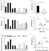
Hemolytic effects on human erythrocytes and the production of phospholipase C (CPA) and perfringolysin O (PFO) in culture supernatants of clinical isolates. Five strains in group H and four strains in group NH, except for the two slow-growing strains (NH5 and NH6), were studied. Strain 13 (St 13), a standard strain, was also examined for comparison. (A) Hemolytic effect of culture supernatants of clinical isolates on a 40% human erythrocyte suspension. The hemolysis rate of each strain was calculated according to a formula. The solid bars show strains in group H, the open bars show strains in group NH, and the diagonal striped bar shows strain 13. The values are the average of three independent experiments conducted with technical triplicates, and the error bars indicate the SDs. (B) Hemolytic effect of group H (n = 5) and group NH (n = 4) strains on a 40% human erythrocyte suspension. *p < 0.05 (C) CPA concentration in culture supernatants of clinical isolates. The values are the average of three independent experiments conducted with technical duplicates, and the error bars indicate the SDs. (D) Correlation between the hemolysis rate and CPA production. The hemolysis rate is plotted against the CPA concentration. (E) Expression of PFO in culture supernatants of clinical isolates, as determined by Western blot analysis. Relative PFO expression normalized to that of strain 13 is expressed numerically. (F) Correlation between the hemolysis rate and PFO production. The hemolysis rate is plotted against the relative PFO expression level normalized to that of strain 13.
Figure 3
Figure 3
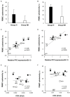
Cytotoxicity of culture supernatants of clinical isolates in human peripheral blood mononuclear cells (PBMCs). (A) Cytotoxicity of 1% culture supernatants from group H (n = 5) and group NH (n = 4) strains in human PBMCs for 4 h. (B) Cytotoxicity of 5% culture supernatant in human PBMCs for 4 h. The values are the average of five independent experiments conducted with technical triplicates, and the error bars indicate the SDs. (C) Correlation between cytotoxicity in human PBMCs and PFO production in 1% culture supernatants. The average cytotoxicity in PBMCs from five healthy donors is plotted against the relative PFO expression normalized to that of strain 13. (D) Correlation between cytotoxicity in human PBMCs and PFO production in 5% culture supernatants. (E) Correlation between cytotoxicity in human PBMCs and CPA production in 1% culture supernatants. The average cytotoxicity in PBMCs from five healthy donors is plotted against the CPA concentration. (F) Correlation between cytotoxicity in human PBMCs and CPA production in 5% culture supernatants.
Figure 4
Figure 4
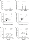
Interleukin-6 (IL-6) and interleukin-8 (IL-8) production by PBMCs induced by supernatants of clinical isolates. Human PBMCs collected from five healthy volunteers on the day of the experiment were stimulated with 1% supernatants of clinical isolates for 4 h. Gifu anaerobic medium (GAM) broth (1%) was used as the control. The graphs show the percentages of IL-6 (A) and IL-8 (B) production normalized to the PBMC count in group H (n = 25) and group NH (n = 20) compared to the control. *p < 0.01 (C) Correlation between IL-6 production and PFO expression. The average amount of IL-6 produced in five healthy donors is plotted against relative PFO expression normalized to that of strain 13. (D) Correlation between IL-8 production and PFO production. The average amount of IL-8 produced in five healthy donors is plotted against relative PFO expression normalized to that of strain 13. (E) Correlation between IL-6 production and CPA production. The average amount of IL-6 produced in five healthy donors is plotted against CPA production. (F) Correlation between IL-8 production and CPA production. The average amount of IL-8 produced in five healthy donors is plotted against CPA production.
Figure 5
Figure 5
Effects of recombinant PFO (rPFO) and recombinant CPA (rCPA) on human blood cells. The effects of rPFO and rCPA were measured using human blood cells collected from five healthy volunteers on the day of the experiment. (A) Hemolytic effect of rPFO on a 40% human erythrocyte suspension. (B) Hemolytic effect of rCPA on a 40% human erythrocyte suspension. (C) Cytotoxicity of rPFO in human PBMCs. (D) Cytotoxicity of rCPA in human PBMCs. (E) IL-6 production by human PBMCs induced by rPFO. (F) IL-6 production by human PBMCs induced by rCPA. (G) IL-8 production by human PBMCs induced by rPFO. (H) IL-8 production by human PBMCs induced by rCPA. The values are the average of five independent experiments conducted with technical duplicates, and the error bars indicate the SDs.
Figure 6
Figure 6
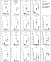
Effects of rPFO and rCPA on human blood cells and the production of various cytokines. The effects of rPFO (63 ng/ml) and rCPA (0.016 U/ml) on human blood cells were evaluated using blood samples from five healthy volunteers. The same volume of phosphate buffered saline (PBS) was used as the control. The black bars indicate the average of five independent experiments conducted with technical duplicates, and the error bars indicate the SDs. **p < 0.01 and *p < 0.05.

References

    1. Aisiku I. P., Yamal J. M., Doshi P., Benoit J. S., Gopinath S., Goodman J. C., et al. . (2016). Plasma cytokines IL-6, IL-8, and IL-10 are associated with the development of acute respiratory distress syndrome in patients with severe traumatic brain injury. Crit. Care 20:288. doi: 10.1186/s13054-016-1470-7, PMID: - DOI - PMC - PubMed
    1. Awad M. M., Ellemor D. M., Boyd R. L., Emmins J. J., Rood J. I. (2001). Synergistic effects of α-toxin and perfringolysin O in Clostridium perfringens-mediated gas gangrene. Infect. Immun. 69, 7904–7910. doi: 10.1128/IAI.69.12.7904-7910.2001, PMID: - DOI - PMC - PubMed
    1. Bryant A. E., Bergstrom R., Zimmerman G. A., Salyer J. L., Hill H. R., Tweten R. K., et al. . (1993). Clostridium perfringens invasiveness is enhanced by effects of theta toxin upon PMNL structure and function: the roles of leukocytotoxicity and expression of CD11/CD18 adherence glycoprotein. FEMS Immunol. Med. Microbiol. 7, 321–336. doi: 10.1111/j.1574-695X.1993.tb00414.x, PMID: - DOI - PubMed
    1. Chinen K. (2020). Sudden death caused by Clostridium perfringens sepsis presenting as massive intravascular hemolysis. Autops. Case Rep. 10:e2020185. doi: 10.4322/acr.2020.185, PMID: - DOI - PMC - PubMed
    1. Hübl W., Mostbeck B., Hartleb H., Pointner H., Kofler K., Bayer P. M. (1993). Investigation of the pathogenesis of massive hemolysis in a case of Clostridium perfringens septicemia. Ann. Hematol. 67, 145–147. doi: 10.1007/BF01701741, PMID: - DOI - PubMed

LinkOut - more resources