. 2021 Sep;22(3):650.
doi: 10.3892/ol.2021.12911. Epub 2021 Jul 9.

A pancancer overview of FBN1, asprosin and its cognate receptor OR4M1 with detailed expression profiling in ovarian cancer

Affiliations

A pancancer overview of FBN1, asprosin and its cognate receptor OR4M1 with detailed expression profiling in ovarian cancer

Rachel Kerslake et al. Oncol Lett. 2021 Sep.

Abstract

Ovarian cancer affects >295,000 women worldwide and is the most lethal of gynaecological malignancies. Often diagnosed at a late stage, current research efforts seek to further the molecular understanding of its aetiopathogenesis and the development of novel biomarkers. The present study investigated the expression levels of the glucogenic hormone asprosin [encoded by fibrillin-1 (FBN1)], and its cognate receptor, olfactory receptor 4M1 (OR4M1), in ovarian cancer. A blend of in silico open access The Cancer Genome Atlas data, as well as in vitro reverse transcription-quantitative PCR (RT-qPCR), immunohistochemistry and immunofluorescence data were used. RT-qPCR revealed expression levels of OR4M1 and FBN1 in clinical samples and in ovarian cancer cell lines (SKOV-3, PEO1, PEO4 and MDAH-2774), as well as the normal human ovarian surface epithelial cell line (HOSEpiC). Immunohistochemical staining of a tissue microarray was used to identify the expression levels of OR4M1 and asprosin in ovarian cancer samples of varying histological subtype and grade, including clear cell carcinoma, serous ovarian cancer and mucinous adenocarcinoma. Immunofluorescence analysis revealed asprosin expression in SKOV-3 and HOSEpiC cells. These results demonstrated the expression of both asprosin and OR4M1 in normal and malignant human ovarian tissues. This research invokes further investigation to advance the understanding of the role of asprosin and OR4M1 within the ovarian tumour microenvironment.

Keywords: asprosin; cancer; fibrillin-1; olfactory receptor; olfactory receptor 4M1; ovarian cancer.


Conflict of interest statement

The authors declare that they have no competing interests.

Figures

Figure 1.
Figure 1.
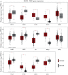
Gene expression of FBN1 in normal tissues. (A) Expression levels of FBN1 in normal human tissues based on available data from The Genotype Tissue Expression project. (B) Co-expression of FBN1 and furin, the enzyme which proteolytically cleaves profibrillin-1 to fibrillin-1 and asprosin, in normal human tissues. The different colours adjacent to furin and FBN1 denote the expression levels of both genes in (B). The darker the colour (dark blue) the higher the expression and the lighter (yellow) the lower the expression (indicated as TPM). The fine different coloured lines underneath the co-expression data in (B) are used for identification purposes and relate to the different coloured violin plots in A. FBN1, fibrillin-1; TPM, transcripts per million.
Figure 2.
Figure 2.
Pancancer profiling of FBN1 expression. Cancer types with significant differences compared with normal tissues (*P<0.01) are presented in the graphs [cancer (red) and normal (grey)]. Cancers with lower expression levels of FBN1 compared with normal samples included UCS, UCEC, THCA, OV, LUSC, LUAD, CESC and BLCA. Those with higher FBN1 expression levels included THYM, STAD, PAAD, HNSC, DLBC and CHOL. BLCA, bladder urothelial carcinoma; CESC, cervical squamous cell carcinoma and endocervical adenocarcinoma; CHOL, cholangiocarcinoma; DLBC, lymphoid neoplasm diffuse large b-cell lymphoma; FBN1, fibrillin-1; GEPIA, Gene Expression Profiling Interactive Analysis; HNSC, head and neck squamous cell carcinoma; LUAD, lung adenocarcinoma; LUSC, lung squamous cell carcinoma; OV, ovarian serous cystadenocarcinoma; PAAD, pancreatic adenocarcinoma; STAD, stomach adenocarcinoma; THCA, thyroid carcinoma; THYM, thymoma; TPM, transcripts per million; UCEC, uterine corpus endometrial carcinoma; UCS, uterine carcinosarcoma.
Figure 3.
Figure 3.
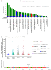
Mutational profile of FBN1. (A) Pancancer overview of the frequency of FBN1 mutations. (B) Copy number of FBN1 alterations across all cancer types (as in Fig. 1A). (C) Location of FBN1 mutations, each lollipop represents an ovarian cancer patient and the corresponding location of the mutation within the gene (Ch15q21.1). Missense mutations are presented as green lollipops, nonsense mutations as black lollipops and splice as orange (source, cBioPortal). CNA, copy number alteration; GISTIC, Genomic Identification of Significant Targets in Cancer; FBN1, fibrillin-1; VUS, variants of unknown significance; RSEM, RNA sequencing by expectation-maximization.
Figure 4.
Figure 4.
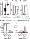
Gene expression of FBN1 and OR4M1 at the ovarian level. (A) Expression data of FBN1 in OC from GEPIA for use as comparison. *P<0.05. Relative expression levels of FBN1 and OR4M1 in OC (red) and normal ovarian tissues (grey) were determined using reverse transcription-quantitative PCR. (B) Significantly lower expression levels of FBN1 in OC samples (OC, n=12; stage III and IV) compared with FBN1 expression in normal ovarian tissue samples from healthy volunteers (n=6). **P<0.001 (samples obtained for the present study; different from the GEPIA cohort in A). (C) Significantly higher expression levels of OR4M1 in OC samples (OC, n=12; stage III and IV) compared with OR4M1 expression in normal ovarian tissue samples from healthy volunteers (n=6). ***P<0.0001. (D) Higher relative expression levels of FBN1 in normal ovarian surface epithelial cells (OSE), and lower expression levels in the studied human ovarian cancer cell lines (SKOV-3, PEO1, PEO4 and MDAH-2774). ***P<0.0001. (E) Lower relative expression levels of OR4M1 in normal ovarian epithelial cells, as well as in the PEO1 and MDAH-2774 human ovarian cancer cell lines, compared with the relatively higher OR4M1 expression noted in SKOV-3 and PEO4 cells. RQ indicates relative change in fold expression to the calibrator gene YWHAZ. FBN1, fibrillin-1; GEPIA, Gene Expression Profiling Interactive Analysis; OC, ovarian cancer; OR4M1, olfactory receptor 4M1; OSE, HOSEpiC cells; TPM, transcripts per million; num(T), number of patients for tumour group; num(N), number of patients for normal group; RQ, relative quantity.
Figure 5.
Figure 5.
Kaplan-Meier plots revealing the prognostic effects of FBN1 expression in OC. (A) OS in OC. (B) OS in early-stage (I and II) OC. (C) OS in late-stage (III and IV) OC. (D) PFS in OC. (E) PFS in early-stage (I and II) OC. (F) PFS in late-stage (III and IV) OC. FBN1, fibrillin-1; HR, hazard ratio; OC, ovarian cancer; OS, overall survival; PFS, progression-free survival.
Figure 6.
Figure 6.
Ovarian tissue microarray, including 90 ovarian cancer cores, stained with asprosin antibody (1:200). Corresponding values for scoring: 0, <10%; 1, 10–25%; 2, 26–50%; 3, 51–75%; and 4, >76% of cells stained. (A) Asprosin staining by histological subtype/grade: LGSC, HGSC, MAC, EAC and CCC. (B) Asprosin staining of early (I and II) and late (III and IV) ovarian cancer stages, revealing no significant difference. (C) HGSC, stage II at ×5 (left) and ×20 (right) magnification. (D) HGSC, stage I at ×5 (left) and ×20 (right) magnification. (E) HGSC, stage III at ×5 (left) and ×20 (right) magnification. CCC, clear cell carcinoma; EAC, endometroid adenocarcinoma; HGSC, high grade serous carcinoma; LGSC, low grade serous carcinoma; MAC, mucinous adenocarcinoma.
Figure 7.
Figure 7.
Immunohistochemical staining of an ovarian tissue microarray, including 90 ovarian cancer cores, with OR4M1 antibody (1:100). (A) OR4M1 staining by histological subtype/grade: LGSC, HGSC, MAC, EAC and CCC. (B) Higher OR4M1 staining in early (I and II) compared with late (III and IV) ovarian cancer stages. *P=0.04. (C) HGSC, stage II at ×5 (left) and ×20 (right) magnification. (D) HGSC, stage I at ×5 (left) and ×20 (right) magnification. (E) HGSC, stage III at ×5 (left) and ×20 (right) magnification. CCC, clear cell carcinoma; EAC, endometroid adenocarcinoma; HGSC, high grade serous carcinoma; LGSC, low grade serous carcinoma; MAC, mucinous adenocarcinoma; NAT, normal adjacent tissue; OR4M1, olfactory receptor 4M1.
Figure 8.
Figure 8.
Immunofluorescence imaging of OSE normal human ovarian epithelial cells and SKOV-3 human serous ovarian cancer cells, with DAPI nuclear staining (red) and with ASP and OR4M1 (green). Magnification, ×100 using a Leica DM4000 microscope (Scale bar, 10 µm). ASP, asprosin; OR4M1, olfactory receptor 4M1.

Similar articles

Cited by

References

    1. Lee B, Godfrey M, Vitale E, Hori H, Mattei MG, Sarfarazi M, Tsipouras P, Ramirez F, Hollister DW. Linkage of Marfan syndrome and a phenotypically related disorder to two different fibrillin genes. Nature. 1991;352:330–334. doi: 10.1038/352330a0. - DOI - PubMed
    1. Klimstra WB, Heidner HW, Johnston RE. The furin protease cleavage recognition sequence of Sindbis virus PE2 can mediate virion attachment to cell surface heparan sulfate. J Virol. 1999;73:6299–6306. doi: 10.1128/JVI.73.8.6299-6306.1999. - DOI - PMC - PubMed
    1. Romere C, Duerrschmid C, Bournat J, Constable P, Jain M, Xia F, Saha PK, Del Solar M, Zhu B, York B, et al. Asprosin, a Fasting-Induced Glucogenic Protein Hormone. Cell. 2016;165:566–579. doi: 10.1016/j.cell.2016.02.063. - DOI - PMC - PubMed
    1. O'Neill B, Simha V, Kotha V, Garg A. Body fat distribution and metabolic variables in patients with neonatal progeroid syndrome. Am J Med Genet A. 2007;143A:1421–1430. doi: 10.1002/ajmg.a.31840. - DOI - PubMed
    1. Wang Y, Qu H, Xiong X, Qiu Y, Liao Y, Chen Y, Zheng Y, Zheng H. Plasma Asprosin Concentrations Are Increased in Individuals with Glucose Dysregulation and Correlated with Insulin Resistance and First-Phase Insulin Secretion. Mediators Inflamm. 2018;2018:9471583. doi: 10.1155/2018/9471583. - DOI - PMC - PubMed