Case Reports
. 2021 Jul 17;16(9):2824-2833.
doi: 10.1016/j.radcr.2021.06.025. eCollection 2021 Sep.

Imaging diagnosis of plexiform neurofibroma- unravelling the confounding features: A report of two cases

Affiliations
Case Reports

Imaging diagnosis of plexiform neurofibroma- unravelling the confounding features: A report of two cases

Dr Shabnam Bhandari Grover et al. Radiol Case Rep. .

Abstract

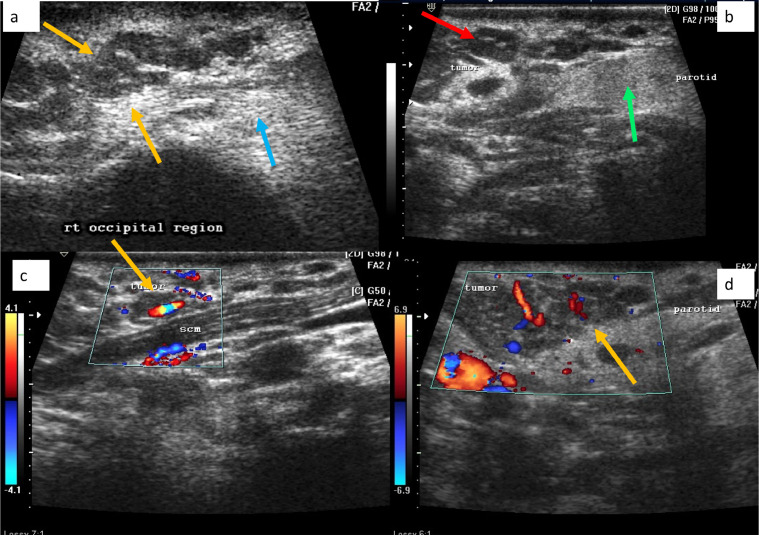
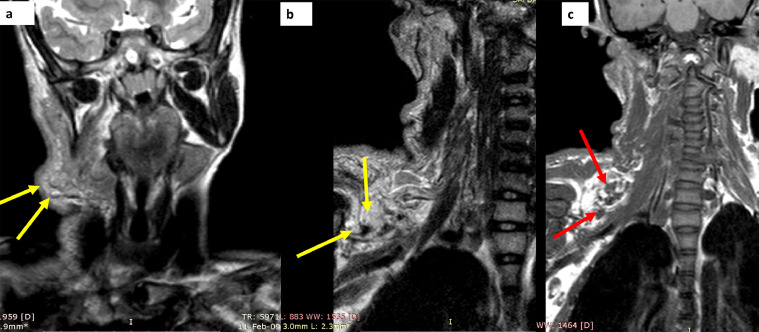
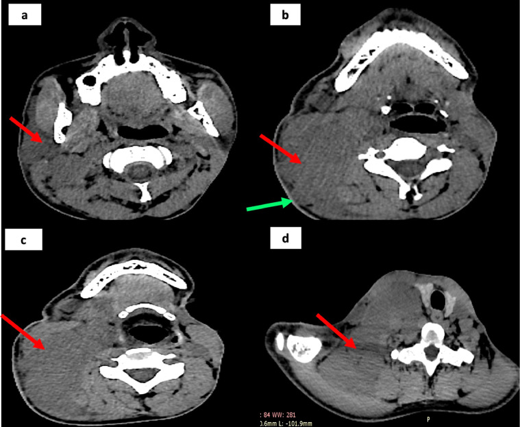
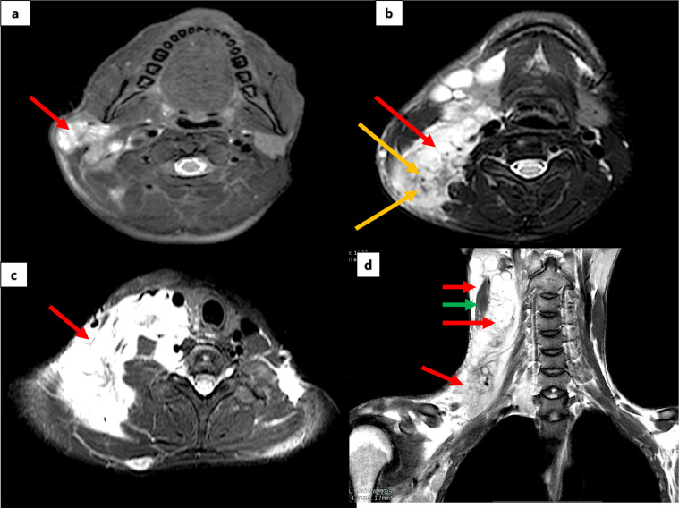
Peripheral nerve sheath tumors such as neurofibroma, comprise 5% of all benign soft tissue tumors and usually occur due to an underlying neurofibromatosis. A plexiform neurofibroma, which is a tumor occurring exclusively in neurofibromatosis1, is a rare entity and is an uncommon variant of neurofibroma. We report the clinical and imaging features of plexiform neurofibroma in two young male patients, in whom the imaging diagnosis was confirmed after biopsy. The report not only aims to highlight the characteristic imaging features of plexiform neurofibroma but we also emphasize the ultrasound appearances which are significantly characteristic and can effectively lead to the correct diagnosis at the preliminary stage of investigation. The tumors which originate from nerve sheath, are large, lobulated masses and demonstrate typical imaging features of simultaneous involvement of subcutaneous and cutaneous tissues along with infiltrative invasion of deeper structures. The tumors characteristically display fat and fluid contents and a "target sign' on evaluation by ultrasound, CT and MRI. Imaging plays an important role in confirming the diagnosis, delineating involved structures, excluding simulating conditions and forewarning a possible malignant transformation.

Keywords: CT; MRI; Neurofibromatosis; Peripheral nerve sheath tumor; Plexiform neurofibroma; Target sign; Ultrasound.


Figures

Fig 1
Fig. 1
Clinical appearance of patient number 1 (a boy of 14 y), shows excessive hirsutism on the right facial and neck regions (white arrows in A, B), with café au lait pigmentation over the preauricular, facial and shoulder regions on the right side (blue arrows in a, red arrows in C). The manubrium is protuberant (green arrow in B) (Color version of figure is available online.)
Fig 2
Fig. 2
Ultrasound of right head and neck region in patient no 1, shows an ill-defined hypo-echoeic tumor, comprising of tubular, serpiginous foci (yellow arrow in A), surrounded by an echogenic background (blue arrow in A), located in the cutaneous and subcutaneous regions. The tumor was abutting the parotid gland (green arrow in B) and a target sign was seen (red arrow in B). Color Doppler evaluation revealed good vascularity within the tumor (yellow arrows in C, D) (Color version of figure is available online.)
Fig 3
Fig. 3
CT scan of the skull base, head and neck region, in patient no 1, shows a lobulated, hypodense tumor in the occipital, facial and neck regions, which is largely cutaneous and subcutaneous in location (yellow arrow in a, green arrows in B). The tumor at the thoracic inlet, insinuates behind the clavicle (red arrow in C). the sternum and clavicle show modelling deformities (white arrows in D, green arrows in E) (Color version of figure is available online.)
Fig 4
Fig. 4
MRI study in patient no 1, obtained as T2 W sequences in the neck and brachial plexus region, shows a homogenously hyper-intense, lobulated tumor over the occipital, submandibular and cervical region (yellow arrows in A, B). the tumor is both superficial and deep to the sternocleidomastoid and shows a target sign in (green arrows in B). The tumor appears to be arising from the C3 to C5 nerve roots (red arrows in C) (Color version of figure is available online.)
Fig 5
Fig. 5
MRI study in patient no 1, obtained as T2 W sequences in the neck and brachial plexus region, shows a homogenously hyper-intense, lobulated tumor over the occipital, submandibular and cervical regions, which is largely subcutaneous and cutaneous in location. The tumor has multiple spherical foci which display a target sign (yellow arrows in A, B). The tumor is hypointense to muscle on T1W sequences (red arrows in C) (Color version of figure is available online.)
Fig 6
Fig. 6
Biopsy image low power (A) and high power (B) of patient number 1, using H and E stain shows nerve bundles (blue arrows) surrounded by collagenous tissue (black arrow). Multiple tightly packed serpentine nuclei are also seen (green arrows). Conforming the diagnosis of plexiform neurofibroma (Color version of figure is available online.)
Fig 7
Fig. 7
Ultrasound examination over the head and neck region in patient no2, shows a multilobulated tumor comprising of spherical and geographic hypoechoeic foci (red arrows in A, B) on a hyperechoeic background (blue arrow in A). The tumor is deep to sternocleidomastoid (green arrow in B) (Color version of figure is available online.)
Fig 8
Fig. 8
NCCT of head, neck and upper thorax in patient number 2, shows a homogenously hypodense tumor, located in the parotid, oropharyngeal, para-pharyngeal, thoracic inlet regions (red arrows in A, B, C and D). the tumor has a lobulated surface (green arrows in B). it displaces the thyroid and trachea across the midline (D) (Color version of figure is available online.)
Fig 9
Fig. 9
MRI examination of patient number 2, over the head, neck and brachial plexus region, using T2W sequences, shows a large homogenously hyper-intense tumor occupying the examined areas, in the superficial as well as deeper planes (red arrows in A, B, C, D). A target sign is visible in B (yellow arrows). The tumor is both superficial and deep to sternocleidomastoid (green arrow in D) (Color version of figure is available online.)
Fig 10
Fig. 10
MRI examination of patient number 2, over the head, neck and brachial plexus region, using STIR sequences, shows a large homogenously hyper-intense tumor in the superficial as well as deeper planes of the examined areas and extending into the axilla (red arrows in A, B). A target sign is seen in the supraclavicular region (green arrows in A) (Color version of figure is available online.)
Fig 11
Fig. 11
Biopsy images of patient no 2, using H and E stain, the low power (A), high power view (B, C) shows: serpentine nuclei marked with green arrow (in B), with a collagenous background seen around the nerve bundles (green arrows in C). Appearances are consistent with neurofibroma (Color version of figure is available online.)

References

    1. Yilmaz S, Ozolek JA, Zammerilla LL, Fitz CR, Grunwaldt LJ, Crowley JJ. Neurofibromas with imaging characteristics resembling vascular anomalies. AJR Am J Roentgenol. 2014;203(6):W697–W705. doi: 10.2214/AJR.13.12409. PMID: 25415736. - DOI - PubMed
    1. Messersmith L, Krauland K. Neurofibroma. 2020 Aug 10. In: StatPearls [Internet]. Treasure Island (FL): StatPearls Publishing; 2020 Jan. PMID: 30969529.
    1. Wise JB, Cryer JE, Belasco JB, Jacobs I, Elden L. Management of head and neck plexiform neurofibromas in pediatric patients with neurofibromatosis type 1. Arch Otolaryngol Head Neck Surg. 2005;131(8):712–718. doi: 10.1001/archotol.131.8.712. PMID: 16103304. - DOI - PubMed
    1. Le C, Bedocs PM. Neurofibromatosis. 2020 Aug 10. In: StatPearls [Internet]. Treasure Island (FL): StatPearls Publishing; 2021 Jan–. PMID: 29083784.
    1. Becker B, Strowd RE., 3rd. Phakomatoses. Dermatol Clin. 2019;37(4):583–606. doi: 10.1016/j.det.2019.05.015. PMID: 31466597. - DOI - PubMed

Publication types